 Login/Register


- Clone
- 5E8 (See other available formats)
- Regulatory Status
- RUO
- Other Names
- CC CKR3, MIP1-alpha receptor like-2, eotaxin receptor, CD193, CCR3
- Isotype
- Mouse IgG2b, κ
- Ave. Rating
- Submit a Review
- Product Citations
- publications

-

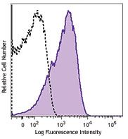
Human peripheral blood leukocytes were stained with CD16 APC and CD193 (clone 5E8) PE (top) or mouse IgG2b, κ PE isotype control (bottom). Data shown was gated on the granulocyte population. -

| Cat # | Size | Price | Quantity Check Availability | Save | ||
|---|---|---|---|---|---|---|
| 310705 | 25 tests | $134 | ||||
| 310706 | 100 tests | $304 | ||||
CD193, also known as CC-chemokine receptor 3 (CCR3), CC CKR3, MIP1-alpha receptor like-2, and eotaxin receptor, is a member of the G protein-coupled seven transmembrane receptors family. It binds to the CC chemokines eotaxin, eotaxin-2, and eotaxin-3 with high affinity. CCR3 has also been reported to bind RANTES, MCP-3, and MCP-4 with low affinity. CCR3 receptor is expressed on human eosinophils, basophils, mast cells, mononuclear phagocytes, platelets, CD34+ hematopoietic progenitor cells, Th2-like lymphocytes, and keratinocytes. CCR3 is thought to play a role in allergic diseases such as bronchial asthma and allergic rhinitis. CCR3 is a co-receptor for HIV-1 and HIV-2, and the binding of eotaxin with CCR3 has been shown to inhibit HIV infection in some cell types.
Product DetailsProduct Details
- Verified Reactivity
- Human
- Antibody Type
- Monoclonal
- Host Species
- Mouse
- Formulation
- Phosphate-buffered solution, pH 7.2, containing 0.09% sodium azide and BSA (origin USA)
- Preparation
- The antibody was purified by affinity chromatography, and conjugated with PE under optimal conditions.
- Concentration
- Lot-specific (to obtain lot-specific concentration and expiration, please enter the lot number in our Certificate of Analysis online tool.)
- Storage & Handling
- The antibody solution should be stored undiluted between 2°C and 8°C, and protected from prolonged exposure to light. Do not freeze.
- Application
-
FC - Quality tested
- Recommended Usage
-
Each lot of this antibody is quality control tested by immunofluorescent staining with flow cytometric analysis. For flow cytometric staining, the suggested use of this reagent is 5 µl per million cells in 100 µl staining volume or 5 µl per 100 µl of whole blood.
- Excitation Laser
-
Blue Laser (488 nm)
Green Laser (532 nm)/Yellow-Green Laser (561 nm)
- Application Notes
-
Additional reported applications (for the relevant formats) include: The 5E8 antibody is useful for immunofluorescent staining and flow cytometric analysis of CCR3 expression.
It has been observed that the 5E8 antibody clone can interact with PE/Cyanine7 antibody conjugates during multi-color staining, potentially leading to unwanted staining. This interaction can be resolved by sequentially staining with the 5E8 antibody first and then followed by the PE/Cyanine7 conjugate of interest. -
Application References
(PubMed link indicates BioLegend citation) -
- Beauvillian C, et al. 2011. Blood 117:1196. PubMed
- Product Citations
-
- RRID
-
AB_345395 (BioLegend Cat. No. 310705)
AB_345396 (BioLegend Cat. No. 310706)
Antigen Details
- Structure
- G-protein coupled seven transmembrane domain receptor, 356 amino acids
- Distribution
-
Eosinophils, basophils, mast, mononuclear phagocytes, platelets, CD34+ hematopoietic progenitor, Th2, keratinocytes
- Function
- Co-receptor for HIV-1 and HIV-2, allergy
- Receptors
- Eotaxin, eotaxin-2, eotaxin-3
- Cell Type
- Eosinophils, Erythrocytes, Hematopoietic stem and progenitors, Macrophages, Mast cells, Thymocytes
- Biology Area
- Immunology
- Molecular Family
- CD Molecules, Cytokine/Chemokine Receptors, GPCR
- Antigen References
-
1. Gerard W, et al. 1996. J. Exp. Med. 183:2437.
2. Uguccioni C, et al. 1997. J. Clin. Invest. 100:1137.
3. Sallusto F, et al. 1997. Science. 277:2005.
4. Loetscher P, et al. 2001. J. Biol. Chem. 276:2986. - Regulation
- Upregulated by high affinity Fc IgE receptor ligation (mast cells), RANTES (keratinocytes), IFNg (monocytes, neutrophils), HIV tat protein (basophils), IL-3, IL-5 and GM-CSF (CD34+ progenitor cells), IL-2 and IL-4(T cells). Downregulated by IL-
- Gene ID
- 1232 View all products for this Gene ID
- UniProt
- View information about CD193 on UniProt.org
Related Pages & Pathways
Pathways


Related FAQs
- What type of PE do you use in your conjugates?
- We use R-PE in our conjugates.
- Does staining at room temperature or even at 37°C help for checking chemokine receptors expression?
-
Due to continuous recycling of many chemokine receptors, it may be worthwhile to consider staining at room temperature or at 37°C if the staining at lower temperature (which can potentially reduce receptor turnover) is not optimal.
Other Formats
View All CD193 Reagents Request Custom ConjugationCustomers Also Purchased
Compare Data Across All Formats
This data display is provided for general comparisons between formats.
Your actual data may vary due to variations in samples, target cells, instruments and their settings, staining conditions, and other factors.
If you need assistance with selecting the best format contact our expert technical support team.
-
Purified anti-human CD193 (CCR3)

Human peripheral blood granulocytes stained with 5E8 PE and ... 
Human paraffin-embedded skin tissue slices were prepared wit... -
PE anti-human CD193 (CCR3)

Human peripheral blood leukocytes were stained with CD16 APC... 
-
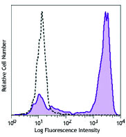
Brilliant Violet 605™ anti-human CD193 (CCR3)

Human peripheral blood leukocytes were stained with CD16 APC... 
-
APC anti-human CD193 (CCR3)
-
Alexa Fluor® 647 anti-human CD193 (CCR3)

Human peripheral blood granulocytes stained with CD16 FITC a... -
APC/Cyanine7 anti-human CD193 (CCR3)

Human peripheral blood leukocytes were stained with CD16 FIT... -
Brilliant Violet 421™ anti-human CD193 (CCR3)

Human peripheral blood leukocytes were stained with CD16 FIT... 
-
PerCP/Cyanine5.5 anti-human CD193 (CCR3)

Human peripheral blood granulocytes were stained with CD16 A... -
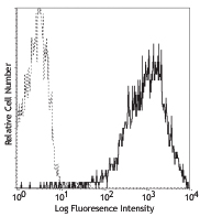
FITC anti-human CD193 (CCR3)

Human peripheral blood leukocytes were stained with CD16 APC... 
-
Brilliant Violet 510™ anti-human CD193 (CCR3)

Human peripheral blood leukocytes were stained with CD16 FIT... 
-
Biotin anti-human CD193 (CCR3)

Human peripheral blood leukocytes were stained with CD16 APC... 
-
APC/Fire™ 750 anti-human CD193 (CCR3)

Human peripheral blood leukocytes were stained with CD16 FIT... 
-
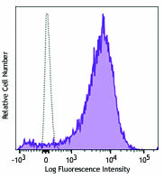
PE/Dazzle™ 594 anti-human CD193 (CCR3)

Human peripheral blood leukocytes were stained with CD16 FIT... 
-
TotalSeq™-A0397 anti-human CD193 (CCR3)
-
TotalSeq™-C0397 anti-human CD193 (CCR3)
-
Brilliant Violet 711™ anti-human CD193 (CCR3) Antibody

Human peripheral blood leukocytes were stained with CD16 APC... -
TotalSeq™-D0397 anti-human CD193 (CCR3)
-
TotalSeq™-B0397 anti-human CD193 (CCR3)
-
Spark Blue™ 550 anti-human CD193 (CCR3) (Flexi-Fluor™)
-
Spark Red™ 718 anti-human CD193 (CCR3) (Flexi-Fluor™)


Follow Us