 Login/Register


- Regulatory Status
- RUO
- Other Names
- Mitochondrial labeling, DiIC1(5)
- Ave. Rating
- Submit a Review
- Product Citations
- publications

-

HeLa cells were stained with 0.25µM CytoPhase™ Violet dye (red) for 60 minutes at 37°C. Then 20 nM of MitoSpy™ NIR DiIC1(5) (green) was added for an additional 30 minutes at 37°C. The Z-stack images were captured on an LSM 880 with Airyscan using a 63x Oil objective. The files were Airyscan processed and a Maximum Intensity projection was created using Zen software; all courtesy of Biophotonics Core Facility at Salk Institute. -

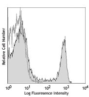
Human T-cell leukemia cell line, Jurkat, was treated (left) or un-treated (right) with 1.0 µg/ml of LEAF™ purified anti-human CD95. At the end of incubation, cells were washed twice, (once with PBS then second time with Annexin V Binding Buffer), then were stained with 5 nM of MitoSpy™ NIR DilC1(5), Brilliant Violet 421™ Annexin V, and Helix NP™ Green (Cat. No. 425303 at 1.25 nM) for 15 minutes at 37°C in Annexin V Binding Buffer (Cat. No. 422201). After incubation, cells were washed and resuspended with Annexin V Binding Buffer. Shown gated on live cells.
| Cat # | Size | Price | Quantity Check Availability | Save | ||
|---|---|---|---|---|---|---|
| 424807 | 200 tests | $59 | ||||
MitoSpy™ mitochondrial localization probes are cell-permeant, fluorogenic chemical reagents that are used for labeling mitochondria in live cells. MitoSpy™ NIR DilC1(5) localizes to the mitochondria based on its membrane potential and is useful to indicate cell health as well as for localization. Only useful on live cells, the use of fixatives is not recommended prior to imaging or flow cytometry.
Product DetailsProduct Details
- Verified Reactivity
- Human, Mouse, Rat, All Species
- Molecular Mass
- 510.45 MW
- Preparation
- The stock solution for MitoSpy™ NIR DilC1(5) is prepared by dissolving the lyophilized probe in dimethyl sulfoxide (DMSO) to make a final concentration of 10uM by adding 500uL of DMSO to each vial.
- Storage & Handling
- Store MitoSpy™ NIR DilC1(5) at -20°C.
- Application
-
ICC/Live cell imaging - Quality tested
FC - Verified - Recommended Usage
-
Each lot of this reagent is quality control tested by immunocytochemistry staining. For immunocytochemistry microscopy, a concentration range of 1 to 50 nM is recommended. For flow cytometric staining, a concentration range of 1 to 50 nM is recommended. It is recommended that the reagent be titrated for optimal performance for each application.
- Application Notes
-
MitoSpy™ NIR DilC1(5) is excited at 638 nm and emits at 658 nm.
Protocol for Live Cell Microscopy- Reconstitute MitoSpy™ NIR DilC1(5) to a 10uM concentration with DMSO by adding 500uL to an individual vial of lyophilized material. Protect the stock solution from light and keep frozen for storage.
- Prepare the working solution for MitoSpy™ NIR DilC1(5) in 37°C pre-warmed culture media, which will vary by cell line and type of imaging required. When labeling mitochondria for live cell imaging a concentration of 1-50 nM is recommended.
- Grow cells to a desired confluency and wash once with pre-warmed 1X PBS.
- Add the working solution of MitoSpy™ NIR DilC1(5) to the live cells and incubate at 37°C for 20-30 minutes.
- Wash the cells twice with warm 1XPBS or culture media.
- Cells are now ready to be imaged live with a fluorescence microscope. MitoSpy™ NIR DilC1(5) is poorly retained with PFA fixation.
Antigen Details
- Distribution
-
Mitochondria
- Biology Area
- Apoptosis/Tumor Suppressors/Cell Death, Cell Biology, Mitochondrial Function, Neuroscience
- Molecular Family
- Mitochondrial Markers
- Gene ID
- NA


Follow Us