 Login/Register


- Regulatory Status
- RUO
- Other Names
- Calcein, Calcien Violet-AM
- Ave. Rating
- Submit a Review
- Product Citations
- publications

-

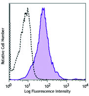
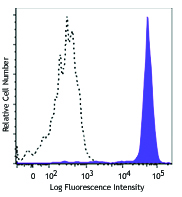
Fresh (top) or day-old C57BL/6 splenocytes (bottom) were stained with 0.01 µM Calcein Violet-AM and a cell-impermeant nucleic acid dye, SYTOX™ Red (colored). Black figure represents unstained splenocytes. -

| Cat # | Size | Price | Quantity Check Availability | Save | ||
|---|---|---|---|---|---|---|
| 425203 | 10 x 25 µg | $211 | ||||
Calcein Violet-AM is a fluorogenic, cell-permeant fluorescent probe that indicates cellular health by detecting the activity of nonspecific esterases. When the acetoxymethyl ester is intact the probe is non-fluorescent, but when it is cleaved by nonspecific esterases present in healthy cells, the probe can be excited and emit at 400nm/452nm respectively. The signal of Calcein Violet-AM is proportional to the cell vitality, as esterase activity decreases in cells with poor vitality. This cell-permeant probe is not retained with fixation and permeabilization.
Product DetailsProduct Details
- Preparation
- Calcein Violet-AM labeling kit consists of lyophilized Calcein Violet-AM and anhydrous DMSO. For reconstitution, bring the kit to room temperature and add 40 µL of DMSO to one vial of Calcein Violet-AM dye until fully dissolved.
- Storage & Handling
- Calcein Violet-AM should be stored at -20°C upon receipt. Do not open vials until needed. Once the DMSO is added to the Calcein Violet-AM, use immediately or store at -20°C in a dry place and protected from light, preferably in a desiccator or in a container with desiccant for no more than one month.
- Application
-
FC - Quality tested
- Recommended Usage
-
This lot has been tested by flow cytometric analysis of cell vitality. It can be used at concentrations ranging from 0.1 – 0.001 µM for cell labeling. It is recommended that the reagent be titrated for optimal performance for each cell type, culturing condition, or application.
- Application Notes
-
The molecular weight of Calcein Violet-AM is 616.48 Da. The maximum excitation and emission wavelengths of Calcein Violet-AM are 400 nm/452 nm, respectively. Each 25 µg of Calcein Violet-AM may be reconstituted with 40 µL of anhydrous DMSO to yield a stock concentration of 1 mM.
Materials Provided:
10 vials x 25 µg Calcein Violet-AM
2 vials of 500 ul anhydrous DMSO
Calcein Violet-AM Labeling Procedure:
1. Prior to reconstitution, spin down the vial of lyophilized reagent in a microcentrofuge to ensure the reagent is at the bottom of the vial.
2. Prepare a 1 mM stock solution by reconstituting 1 vial of lyophilized Calcein Violet-AM dye with 40 µl of anhydrous DMSO.
3. Prepare a 1 µM working solution by diluting 1 µL of 1 mM Calcein Violet-AM stock solution in 1 mL PBS.
4. Spin down cells and adjust the cell suspension to 1x10⁷ cells/mL in PBS.
5. Add 10 µl of the 1 µM Calcein Violet-AM working solution to each mL of cell suspension for a final concentration of 0.01 µM.
6. Incubate cells for 20 minutes at room temperature or at 37°C, protected from light.
7. Pellet cells and resuspend in pre-warmed cell culture medium.
8. Incubate cells for 10 minutes to ensure optimal retention of the Calcein Violet-AM.
9. After incubation, Calcein Violet-AM labeled cells are ready for downstream applications or analysis. -
Application References
(PubMed link indicates BioLegend citation) -
- Eastburn DJ, et al. 2014. Nucleic Acids Res. 42:e128. (FC)
- Zhou Q, et al. 2012. Blood. 120:4334. (FC)
- Fr÷hlich JD, et al. 2012. Am. J. Pathol. 180:153.
- Wang T, et al. 2012. ACS Nano. 6:1251.
- Lis R, et al. 2011. Int. J. Cancer 128:715. (FC)
- Cole LE, et al. 2007. Infect. Immun. 75:4127. (IF)
Antigen Details
- Structure
- Acetoxymethyl ester.
- Distribution
-
Cytoplasmic.
- Function
- Cell vitality indicator.
- Interaction
- Nonspecific esterases.
- Biology Area
- Apoptosis/Tumor Suppressors/Cell Death, Cell Biology, Cell Proliferation and Viability
- Gene ID
- NA


Follow Us