 Login/Register


- Clone
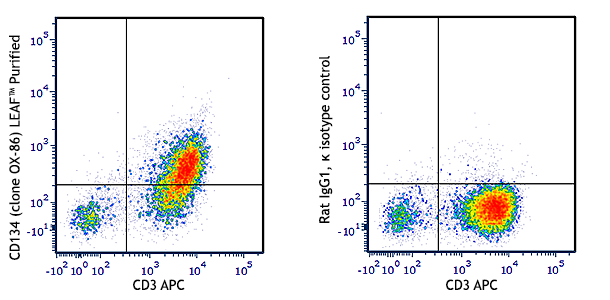
- OX-86 (See other available formats)
- Regulatory Status
- RUO
- Other Names
- TNFRSF4, ACT35, OX-40
- Isotype
- Rat IgG1, κ
- Ave. Rating
- Submit a Review
- Product Citations
- publications

-

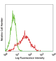
Con-A + IL-2 stimulated (3 days) C57BL/6 splenocytes were stained with CD134 (clone OX-86) Brilliant Violet 711™ (filled histogram) or rat IgG1, κ Brilliant Violet 711™ isotype control (open histogram).
| Cat # | Size | Price | Quantity Check Availability | Save | ||
|---|---|---|---|---|---|---|
| 119421 | 50 µg | $321 | ||||
CD134 is a type I integral membrane protein also known as OX-40, ACT35, and tumor necrosis factor receptor superfamily member 4 (TNFRSF4). This receptor is expressed on activated CD4+ and CD8+ T cells and B cells. The OX-40 receptor binds to the OX-40 ligand (CD252) to provide a costimulatory signal that is independent of CD28. Blockade of OX40-OX40 ligand interactions has been shown to ameliorate experimental EAE and inflammatory bowel disease, which implies that these interactions are important in the pathogenesis of some autoimmune diseases.
Product DetailsProduct Details
- Verified Reactivity
- Mouse
- Antibody Type
- Monoclonal
- Host Species
- Rat
- Immunogen
- Recombinant mouse OX-40-CD4 chimeric protein
- Formulation
- Phosphate-buffered solution, pH 7.2, containing 0.09% sodium azide and BSA (origin USA).
- Preparation
- The antibody was purified by affinity chromatography and conjugated with Brilliant Violet 711™ under optimal conditions.
- Concentration
- 0.2 mg/ml
- Storage & Handling
- The antibody solution should be stored undiluted between 2°C and 8°C, and protected from prolonged exposure to light. Do not freeze.
- Application
-
FC - Quality tested
- Recommended Usage
-
Each lot of this antibody is quality control tested by immunofluorescent staining with flow cytometric analysis. For flow cytometric staining, the suggested use of this reagent is ≤0.75 µg per million cells in 100 µl volume. It is recommended that the reagent be titrated for optimal performance for each application.
Brilliant Violet 711™ excites at 405 nm and emits at 711 nm. The bandpass filter 710/50 nm is recommended for detection, although filter optimization may be required depending on other fluorophores used. Be sure to verify that your cytometer configuration and software setup are appropriate for detecting this channel. Refer to your instrument manual or manufacturer for support. Brilliant Violet 711™ is a trademark of Sirigen Group Ltd.
Learn more about Brilliant Violet™.
This product is subject to proprietary rights of Sirigen Inc. and is made and sold under license from Sirigen Inc. The purchase of this product conveys to the buyer a non-transferable right to use the purchased product for research purposes only. This product may not be resold or incorporated in any manner into another product for resale. Any use for therapeutics or diagnostics is strictly prohibited. This product is covered by U.S. Patent(s), pending patent applications and foreign equivalents. - Excitation Laser
-
Violet Laser (405 nm)
- Application Notes
-
Clone OX-86 has been reported to act as an agonist and stimulate OX-40.
-
Application References
(PubMed link indicates BioLegend citation) -
- Higgins LM, et al. 1999. J. Immunol. 162:486. (FC, IHC)
- Al-Shamkhani A, et al. 1996. Eur. J. Immunol. 26:1695. (Costim)
- del Rio ML, et al. 2011. Transpl. Int. 24:501. (FC) PubMed
- Product Citations
-
- RRID
-
AB_2687176 (BioLegend Cat. No. 119421)
Antigen Details
- Structure
- TNF receptor superfamily, 50 kD
- Distribution
-
Activated CD4+ and CD8+ T cells, activated B cells.
- Function
- Receptor for OX-40 ligand, provides co-stimulatory signal for lymphocyte proliferation independent of CD28; thought to play a role in the pathogenesis of some autoimmune diseases
- Ligand/Receptor
- OX-40 ligand
- Cell Type
- B cells, T cells, Tregs
- Biology Area
- Immunology
- Molecular Family
- CD Molecules, Immune Checkpoint Receptors
- Antigen References
-
1. Al-Shamkhani A, et al. 1996. Eur. J. Immunol. 26:1695.
2. Weinberg AD, et al. 1999. J. Immunol. 162:1818.
3. Akira H, et al. 1999. J. Immunol. 162:7058.
4. Pippig SD, et al. 1999. J. Immunol. 163:6520.
5. Higgins LM, et al. 1999. J. Immunol. 162:486. - Gene ID
- 22163 View all products for this Gene ID
- UniProt
- View information about CD134 on UniProt.org
Related Pages & Pathways
Pathways


Related FAQs
Other Formats
View All CD134 Reagents Request Custom Conjugation| Description | Clone | Applications |
|---|---|---|
| PE anti-mouse CD134 (OX-40) | OX-86 | FC |
| Biotin anti-mouse CD134 (OX-40) | OX-86 | FC |
| Brilliant Violet 421™ anti-mouse CD134 (OX-40) | OX-86 | FC |
| APC anti-mouse CD134 (OX-40) | OX-86 | FC |
| PE/Cyanine7 anti-mouse CD134 (OX-40) | OX-86 | FC |
| PE/Dazzle™ 594 anti-mouse CD134 (OX-40) | OX-86 | FC |
| Brilliant Violet 711™ anti-mouse CD134 (OX-40) | OX-86 | FC |
| Brilliant Violet 605™ anti-mouse CD134 (OX-40) | OX-86 | FC |
| APC/Fire™ 750 anti-mouse CD134 (OX-40) | OX-86 | FC |
| PerCP/Cyanine5.5 anti-mouse CD134 (OX-40) | OX-86 | FC |
| TotalSeq™-A0195 anti-mouse CD134 (OX-40) | OX-86 | PG |
| Ultra-LEAF™ Purified anti-mouse CD134 (OX-40) | OX-86 | FC |
| TotalSeq™-C0195 anti-mouse CD134 (OX-40) | OX-86 | PG |
Customers Also Purchased
Compare Data Across All Formats
This data display is provided for general comparisons between formats.
Your actual data may vary due to variations in samples, target cells, instruments and their settings, staining conditions, and other factors.
If you need assistance with selecting the best format contact our expert technical support team.
-
PE anti-mouse CD134 (OX-40)

Con A-stimulated (3 days) C57BL/6 splenocytes were stained w... -
Biotin anti-mouse CD134 (OX-40)

Con A-stimulated C57BL/6 mouse splenocytes stained with biot... -
Brilliant Violet 421™ anti-mouse CD134 (OX-40)

Con A-stimulated (3 days) C57BL/6 splenocytes were stained w... -
APC anti-mouse CD134 (OX-40)

Con A-stimulated (3 days) C57BL/6 splenocytes were stained w... 
-
PE/Cyanine7 anti-mouse CD134 (OX-40)

C57BL/6 mouse splenocytes were stimulated for three days wit... -
PE/Dazzle™ 594 anti-mouse CD134 (OX-40)

C57BL/6 mouse splenocytes were stimulated for three days wit... -
Brilliant Violet 711™ anti-mouse CD134 (OX-40)

Con-A + IL-2 stimulated (3 days) C57BL/6 splenocytes were st... -
Brilliant Violet 605™ anti-mouse CD134 (OX-40)

Con-A + IL-2 stimulated (3 days) C57BL/6 splenocytes were st... -
APC/Fire™ 750 anti-mouse CD134 (OX-40)

Con-A + IL-2 stimulated (3 days) C57BL/6 splenocytes were st... 
-
PerCP/Cyanine5.5 anti-mouse CD134 (OX-40)

Con-A + IL-2 stimulated (3 days) C57BL/6 splenocytes were st... -
TotalSeq™-A0195 anti-mouse CD134 (OX-40)
-
Ultra-LEAF™ Purified anti-mouse CD134 (OX-40)

Con A-stimulated (3 days) C57BL/6 splenocytes were stained w... -
TotalSeq™-C0195 anti-mouse CD134 (OX-40)


Follow Us