 Login / Register


- Clone
- 150D (See other available formats)
- Regulatory Status
- RUO
- Other Names
- Forkhead box protein P3, Scurfin, JM2, IPEX, Zinc finger protein JM2
- Ave. Rating
- Submit a Review
- Product Citations
- publications

-

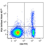
BALB/c mouse splenocytes were stained with True-Nuclear™ Mouse Treg Flow™ Kit (FOXP3 Alexa Fluor® 488/CD4 APC/CD25 PE) -

| Cat # | Size | Price | Quantity Check Availability | Save | ||
|---|---|---|---|---|---|---|
| 320029 | 25 tests | 423€ | ||||
T regulatory (Treg) cells are a subset of T lymphocytes which is characterized by CD4+/CD25+/FOXP3+. These naturally occurring Treg cells originate in the thymus, and comprise 2-10% of peripheral CD4+ T cells. It has been shown that Treg cells are able to inhibit T cells proliferation and cytokine production and play critical roles in preventing autoimmunity as well as in controlling tumor immunity and transplantation tolerance. Impaired Treg function or Treg cell deficiency will develop variety of autoimmune diseases, while higher frequency of Treg cells will cause hypo-immune response to pathogens.
BioLegend's True-Nuclear™ Mouse Treg Flow™ Kit is designed and formulated specifically for immunofluroscence staining and flow cytometric analysis of mouse Treg cells in a mixed lymphocyte population. This kit is composed of fluorochrome conjugated anti-mouse CD4, CD25, FOXP3 antibodies, and the critical buffers. It is easy to use for identification of Treg cells.
Product Details
- Verified Reactivity
- Mouse
- Antibody Type
- Monoclonal
- Concentration
- Lot-specific (to obtain lot-specific concentration and expiration, please enter the lot number in our Certificate of Analysis online tool.)
- Storage & Handling
- This kit is guaranteed for six months. Upon receipt, store between 2°C and 8°C, and protected from prolonged exposure to light. Do not freeze.
- Application
-
ICFC - Quality tested
- Application Notes
-
Materials Provided:
1. Alexa Fluor® 488 anti-mouse FOXP3 - 25 tests
2. Alexa Fluor® 488 Mouse IgG1, ? isotype control - 25 tests
3. True-Nuclear™ Transcription Factor Buffer Set - 120 tests (Cat. No. 424401)
4. anti-mouse CD4 APC/CD25 PE Cocktail - 50 tests
Materials Not Included:
1. Cell Staining Buffer (Cat. No. 420201)
2. Single color compensation controlsImmunofluorescence Staining Procedures:
1. Perform cell surface staining as described in BioLegend's Cell Surface Immunofluorescence Staining Protocol. Add 20 µL of the anti-mouse CD4 APC/CD25 PE cocktail to each tube and incubate in the dark for 20 minutes.
2. Add 2 mL of the cell staining buffer, centrifuge tubes at 400 x g at room temperature for five minutes, and discard the supernatant.
3. Repeat Step 2, for a total of two washes.
4. Add 1 mL of the Transcription Factor 1X Fix solution to each tube, vortex, and incubate at room temperature in the dark for 45-60 minutes.
5. Without washing, add 2 mL of the Transcription Factor 1X Perm Buffer to each tube.
6. Centrifuge tubes at 400 x g at room temperature for five minutes, and discard the supernatant.
7. Add 2 mL of the Transcription Factor 1X Perm Buffer to each tube.
8. Centrifuge tubes at 400 x g at room temperature for five minutes, and discard the supernatant.
9. Resuspend the cell pellet in 100 µl of the Transcription Factor 1XPerm Buffer.
10. Add 5 µL of Alexa Fluor® 488 anti-mouse FOXP3 antibody or 5 µL of Alexa Fluor® 488 mouse IgG1, ? isotype control into the appropriate tubes. Incubate in the dark at room temperature for at least 30 minutes.
11. Add 2 mL of the Transcription Factor 1X Perm Buffer to each tube.
12. Centrifuge tubes at 400 x g at room temperature for five minutes, and discard the supernatant.
13. Add 2 mL of the cell staining buffer.
14. Centrifuge tubes at 400 x g at room temperature for five minutes, and discard the supernatant.
15. Resuspend in 0.5 mL cell staining buffer and then acquire tubes on a flow cytometer.
Caution: The True-Nuclear™ Transcription Factor Buffer Set contains paraformaldehyde, which is toxic and mutagenic. Please handle with caution. Wear gloves, lab coats, and necessary protection to avoid direct contact.
NOTE: For flow cytometric staining with this clone, True-Nuclear™ Transcription Factor Buffer Set (Cat. No. 424401) offers improved staining and is highly recommended over the Foxp3/Perm Buffer Set (Cat. No. 421403). - Application References
-
- Roncador G, et al. 2005 Eur. J. Immunol. 35:1681.
- Mayack. S,et al. 2006. J. Immunol.176:2059. J. Immunol
- Yang ZZ, et al. 2006. Blood 107:3639.
- Gavin MA, et al. 2006. P. Natl. Acad. Sci. USA 103:6659.
- Groh V, et al. 2006. Nature Immunology 7:755.
- Lewkowicz P, et al. 2006 J. Immunol. 177:7155.
- Luke PPW, et al. 2006. Amer. J. Transplant. 6(9):2023.
- Bamias G, et al. 2007. J. Immunol. 178:1809.
- Valencia X, et al. 2007. J. Immunol. 178:2579.PubMed
- Davidson TS, et al. 2007. J. Immunol. 178:4022.
- MacDonald K PA, et al. 2007. Blood doi:10.1182/blood-2007-01-067249.
- Jaffar Z, et al. 2007. J. Immunol. 179:6193.
- Müller M, et al. 2007. J. Immunol. 179:2774.
- Jordan JM,et al. 2008.Infect Human. 76:3717. PubMed
- Golovina TN,et al. 2008. J. Immunol. 181:2855. PubMed
- Fallarino F, et al. 2009. J. Exp Med. 206:2511. PubMed
- Banham Alison, et al. 2009. Vet Immunol and Immunop 127.3-4:376-381
- Klunker S, et al. 2009. J. Exp Med. PubMed
- Haque A, et al. 2010. J. Immunol. 184:2583. PubMed
- Liu Y, et al. 2012. Food Chem Toxicol. 50:1920. PubMed
- Product Citations
-
Antigen Details
- Distribution
-
T regulatory cells
- Cell Type
- Tregs
- Biology Area
- Cell Biology, Immunology, Transcription Factors
- Molecular Family
- CD Molecules, Nuclear Markers
- Antigen References
-
1. Hori S, et al. 2003. Science 299:1057.
2. Fontenot JD, et al. 2003. Nature Immunol. 4:330.
3. Ferguson PJ, et al. 2000. Am. J. Med. Genet. 90:390.
4. Bennett CL, et al. 2001. Nature Genet. 27:20.
5. Allan SE, et al. 2005. J. Clin. Invest. 115:3276. - Gene ID
- 20371 View all products for this Gene ID 317382 View all products for this Gene ID 50943 View all products for this Gene ID


Related FAQs
- What type of PE do you use in your conjugates?
- We use R-PE in our conjugates.
- Can I stain whole blood with anti-FOXP3 using your Foxp3 staining kit?
-
It is not recommended. It is best to use PBMCs for this testing.
- Can FOXP3 be costained with cytokines?
-
The larger holes created by the nuclear permeabilization required for FOXP3 may allow cytokines to leak out of the cell, making it harder to detect lowly-expressed cytokines. You may have to use a control where the cells are only permeabilized through the cell membrane.
Other Formats
View All Reagents Request Custom Conjugation| Description | Clone | Applications |
|---|---|---|
| Purified anti-mouse/rat/human FOXP3 | 150D | WB,ICFC |
| PE anti-mouse/rat/human FOXP3 | 150D | ICFC |
| Alexa Fluor® 488 anti-mouse/rat/human FOXP3 | 150D | ICFC |
| Alexa Fluor® 647 anti-mouse/rat/human FOXP3 | 150D | ICFC |
| True-Nuclear™ Human Treg Flow™ Kit (FOXP3 Alexa Fluor® 488/CD4 PE-Cy5/CD25 PE) | 150D | ICFC |
| True-Nuclear™ Mouse Treg Flow™ Kit (FOXP3 Alexa Fluor® 488/CD4 APC/CD25 PE) | 150D | ICFC |
Customers Also Purchased
Compare Data Across All Formats
This data display is provided for general comparisons between formats.
Your actual data may vary due to variations in samples, target cells, instruments and their settings, staining conditions, and other factors.
If you need assistance with selecting the best format contact our expert technical support team.
-
Purified anti-mouse/rat/human FOXP3

Cell extract from HEK293T cells transfected with either huma... -
PE anti-mouse/rat/human FOXP3

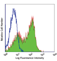
C57BL/6 splenocytes were surface stained with CD4 APC and th... 
-
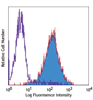
Alexa Fluor® 488 anti-mouse/rat/human FOXP3

C57BL/6 splenocytes were surface stained with CD4 APC and th... 
-
Alexa Fluor® 647 anti-mouse/rat/human FOXP3

C57BL/6 splenocytes were surface stained with CD4 PE and the... 
-
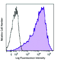
True-Nuclear™ Human Treg Flow™ Kit (FOXP3 Alexa Fluor® 488/CD4 PE-Cy5/CD25 PE)

Human peripheral blood lymphocytes were stained with True-Nu... 
-
True-Nuclear™ Mouse Treg Flow™ Kit (FOXP3 Alexa Fluor® 488/CD4 APC/CD25 PE)

BALB/c mouse splenocytes were stained with True-Nuclear™ Mou... 


Follow Us