 Login / Register


- Clone
- 43D (See other available formats)
- Regulatory Status
- RUO
- Other Names
- Microtubule-associated protein tau, PHF-tau, paired helical filament-tau, neurofibrillary tangle protein, microtubule-associated protein tau, isoform 4, G protein beta1/gamma2 subunit-interacting factor 1
- Previously
-
Covance Catalog# SIG-39402
- Isotype
- Mouse IgG1, κ
- Ave. Rating
- Submit a Review
- Product Citations
- publications

-

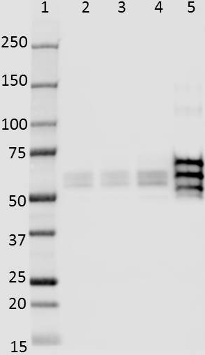
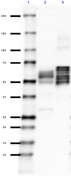
Western blot of purified anti-Tau, 1-100 antibody (clone 43D). Lane 1: Molecular weight marker; Lane 2: 20 µg of normal human brain lysate; Lane 3: 20 µg of Alzheimer's disease human brain lysate. The blot was incubated with 5 µg/mL of the primary antibody overnight at 4°C, followed by incubation with HRP labeled goat anti-mouse IgG (Cat. No. 405306). Enhanced chemiluminescence was used as the detection system. -

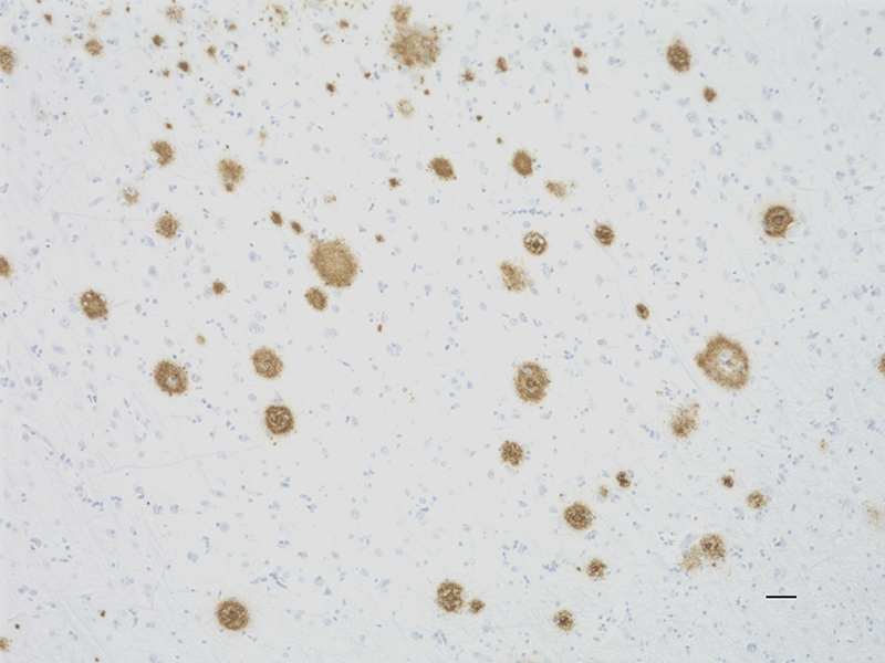
IHC staining of purified anti-Tau, 1-100 antibody (clone 43D) on formalin-fixed paraffin-embedded Alzheimer's disease human brain tissue. Following antigen retrieval using Sodium Citrate H.I.E.R., the tissue was incubated with 1 µg/ml of the primary antibody for 60 minutes at room temperature. BioLegend´s Ultra-Streptavidin (USA) HRP kit (Multi-Species, DAB, Cat. No. 929901) was used for detection followed by hematoxylin counterstaining, according to the protocol provided. The image was captured with a 40X objective. Scale bar: 50 µm
| Cat # | Size | Price | Quantity Check Availability | Save | ||
|---|---|---|---|---|---|---|
| 816603 | 25 µL | 132€ | ||||
| 816601 | 100 µL | 444€ | ||||
Tau protein promotes microtubule assembly and stability. Tau is abundant in neurons of the central nervous system, and is expressed at low levels in astrocytes and oligodendrocytes. Abnormal hyper-phosphorylation, aggregation, and toxic gain of function of tau is associated with several neurological disorders, including Alzheimer’s disease (AD). The major building block of neurofibrillary lesions in AD brains consists of paired helical filaments (PHFs) of abnormally hyperphosphorylated tau. Six isoforms of tau are generated by alternative splicing of the MAPT gene. These isoforms are distinguished by the number of tubulin binding domains, 3 (3R) or 4 (4R), in the C-terminal of the protein and by one (1N), two (2N), or no (0N) inserts in the N-terminal domain. Tau isoforms are differentially expressed during development.
Product DetailsProduct Details
- Verified Reactivity
- Human
- Antibody Type
- Monoclonal
- Host Species
- Mouse
- Formulation
- Phosphate-buffered solution.
- Preparation
- The antibody was purified by affinity chromatography.
- Concentration
- 2 mg/ml
- Storage & Handling
- The antibody solution should be stored undiluted between 2°C and 8°C. Please note the storage condition for this antibody has been changed from -20°C to between 2°C and 8°C. You can also check your vial or your CoA to find the most accurate storage condition for this antibody.
- Application
-
WB - Quality tested
IHC-P - Verified - Recommended Usage
-
Each lot of this antibody is quality control tested by Western blotting. For Western blotting, the suggested use of this reagent is 1.0 - 5.0 µg per ml. For immunohistochemistry, a concentration range of 1.0 - 5.0 µg/ml is suggested. It is recommended that the reagent be titrated for optimal performance for each application.
- Application Notes
-
This antibody is reactive to amino acid residues 1-100 of human Tau. The epitope lies within amino acids 6-18. This clone is specific for all six isoforms of human tau protein.
This antibody clone has been reported for use on IHC of 4% PFA-fixed free floating sections5.
This antibody is exclusively provided by BioLegend. - Product Citations
-
- RRID
-
AB_2715839 (BioLegend Cat. No. 816603)
AB_2564800 (BioLegend Cat. No. 816601)
Antigen Details
- Structure
- Unmodified Tau isoforms have an apparent molecular weight ranging from 33-79 kD. Additional high and low molecular weight Tau species have been observed in brain tissues.
- Distribution
-
Tissue distribution: Central nervous system, peripheral ganglia and nerves, kidney, skeletal, and heart muscle.
Cellular distribution: cytoskeleton, nucleus, plasma membrane and cytosol. - Function
- Tau promotes microtubule assembly and stability. The short tau isoforms allow plasticity of the cytoskeleton whereas the longer isoforms may preferentially play a role in its stabilization.
- Interaction
- Tau interacts with Sequestosome-1, Peptidyl-prolyl cis-trans isomerase FKBP4, Casein kinase I isoform delta, Serine/threonine-protein kinase Sgk1, Laforin, Alpha-synuclein
- Biology Area
- Cell Biology, Neurodegeneration, Neuroscience, Protein Misfolding and Aggregation
- Molecular Family
- Tau
- Antigen References
-
- Augustinack JC, et al. 2002. Acta Neuropath. 103(1):26-35.
- Seubert P, et al. 1995. J Biol Chem. 270(32):18917-22.
- Dong Y, et al. 2012. PLoS One. 7(6):e39386.
- Simic G, et al. 2016. Biomolecules. 6(1):6.
- Dai CL, et al. 2017. Alzheimers Res Ther. 9(1):1. (IHC, WB) [PubMed]
- Baazaoui N, et al. 2017. Alzheimers Res Ther. 9:45. ( WB) [PubMed]
- Dai CL, et al. 2015. J neural Transm (Vienna). 122:607.
- Gene ID
- 4137 View all products for this Gene ID
- UniProt
- View information about Tau 1-100 on UniProt.org


Related FAQs
Other Formats
View All Tau, 1-100 Reagents Request Custom Conjugation| Description | Clone | Applications |
|---|---|---|
| Purified anti-Tau, 1-100 | 43D | WB,IHC-P |
| Alexa Fluor® 594 anti-Tau, 1-100 | 43D | IHC-P |
| Alexa Fluor® 647 anti-Tau, 1-100 | 43D | IHC-P,IHC,WB |
| Alexa Fluor® 488 anti-Tau, 1-100 | 43D | IHC-P,IHC,WB |
| Biotin anti-Tau, 1-100 | 43D | WB,IHC-P,IHC,WB |
Customers Also Purchased
Compare Data Across All Formats
This data display is provided for general comparisons between formats.
Your actual data may vary due to variations in samples, target cells, instruments and their settings, staining conditions, and other factors.
If you need assistance with selecting the best format contact our expert technical support team.
-
Purified anti-Tau, 1-100

Western blot of purified anti-Tau, 1-100 antibody (clone 43D... 
IHC staining of purified anti-Tau, 1-100 antibody (clone 43D... -
Alexa Fluor® 594 anti-Tau, 1-100

Immunofluorescence staining of Alexa Fluor® 594 anti-Tau, 1-... -
Alexa Fluor® 647 anti-Tau, 1-100

IHC staining of Alexa Fluor® 647 anti-Tau, 1-100 antibody (c... -
Alexa Fluor® 488 anti-Tau, 1-100

IHC staining of Alexa Fluor® 488 anti-Tau, 1-100 antibody (c... -
Biotin anti-Tau, 1-100

Western blot of Biotin anti-Tau, 1-100 antibody (clone 43D).... 
IHC staining of Biotin anti-Tau, 1-100 antibody (clone 43D) ...


Follow Us