 Login / Register


- Clone
- 206D (See other available formats)
- Regulatory Status
- RUO
- Other Names
- Forkhead box protein P3, Scurfin, JM2, IPEX, Zinc finger protein JM2
- Isotype
- Mouse IgG1, κ
- Ave. Rating
- Submit a Review
- Product Citations
- publications

-

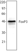
Cell extract from HEK293T cells transfected with human FoxP3 cDNA was resolved by electrophoresis, transferred to nitrocellulose, and probed with monoclonal anti-FoxP3 antibody (clone 206D). Proteins were visualized using a goat anti-mouse secondary conjugated to HRP and a chemiluminescence detection system.
| Cat # | Size | Price | Quantity Check Availability | Save | ||
|---|---|---|---|---|---|---|
| 320101 | 25 µg | 104€ | ||||
| 320102 | 100 µg | 212€ | ||||
FOXP3 is a 50-55 kD transcription factor, also known as Forkhead box protein P3, Scurfin, JM2, or IPEX. It is proposed to be a master regulatory gene and more specific marker of T regulatory cells than most cell surface markers (such as CD4 and CD25). Transduced expression of FOXP3 in CD4+/CD25- cells has been shown to induce GITR, CD103, and CTLA4 and impart a T regulatory cell phenotype. FOXP3 is mutated in X-linked autoimmunity-allergic dysregulation syndrome (XLAAD or IPEX) in humans and in "scurfy" mice. Overexpression of FOXP3 has been shown to lead to a hypoactive immune state suggesting that this transcriptional factor is a central regulator of T cell activity. In human, unlike in mouse, two isoforms of FOXP3 have been reported: one (FOXP3) corresponding to the canonical full-length sequence; the other (FOXP3 δ2) lacking exon 2. The 206D antibody recognizes human FOXP3 epitope in the region of amino acids 105-235.
Product DetailsProduct Details
- Verified Reactivity
- Human
- Reported Reactivity
- Baboon, Cynomolgus, Rhesus, Pigtailed Macaque
- Antibody Type
- Monoclonal
- Host Species
- Mouse
- Immunogen
- Full-length FOXP3 protein
- Formulation
- This antibody is provided in phosphate-buffered solution, pH 7.2, containing 0.09% sodium azide.
- Preparation
- The antibody was purified by affinity chromatography.
- Concentration
- 0.5 mg/ml
- Storage & Handling
- The antibody solution should be stored undiluted between 2°C and 8°C.
- Application
-
WB - Quality tested
ICFC, IHC-F, IHC-P - Reported in the literature, not verified in house - Recommended Usage
-
Each lot of this antibody is quality control tested by immunofluorescent intracellular staining with flow cytometric analysis. For flow cytometric staining, the suggested use of this reagent is ≤ 0.5 µg per 106 cells in 100 µl volume. For Western blotting, the suggested working dilution(s) is ≤ 5.0 µg/ml in antibody dilution buffer. It is recommended that the reagent be titrated for optimal performance for each application.
- Application Notes
-
Additional reported applications (for the relevant formats) include: immunohistochemical staining of acetone-fixed frozen sections1 and formalin-fixed paraffin-embedded sections1,8,19-20, and Western blotting1. The binding of 206D to FOXP3 can be partially blocked by 259D, but 206D does not show significant blocking effect on 259D binding.
NOTE: For flow cytometric staining with this clone, True-Nuclear™ Transcription Factor Buffer Set (Cat. No. 424401) offers improved staining and is highly recommended. - Application References
-
- Roncador G, et al. 2005. Eur. J. Immunol. 35:1681.(IHC)
- Yang ZZ, et al. 2006. Blood 107:3639.
- Liu W, et al. 2006. J. Exp. Med. 203:1701.PubMed
- Bollyky PL, et al. 2007. J. Immunol. 179:744.
- Bell MP, et al. 2007. J. Immunol. 179:1893.
- Tran DQ, et al. 2007. Blood doi:10.1182/blood-2007-06-094656. PubMed
- Gao Q,et al.2007.J Clin Oncol.25:2586.(IHC) PubMed
- Pillai V,et al. 2008. Blood 111:463. PubMed
- Zheng Y, et al. 2008. J. Immunol. 181:1683. PubMed
- Zonios DI, et al. 2008.Blood112:287. PubMed
- Kavanagh B, et al. 2008. Blood. PubMed
- Nevala WK, et al. 2009. Clin Cancer Res. 15:1931. PubMed
- Grant J, et al. 2009. Cytometry B Clin Cytom. 76:69. PubMed
- Nigam P, et al. 2010. J. Immunol. 184:1690. PubMed
- Kmieciak M, et al. 2009. J. Transl. Med. 7:89. (ICFC) PubMed
- Hartigan-O'Connor DJ,et al.2007.J Exp Med.204:2679. PubMed
- Raghaven S, et al. 2009. Ann Rheum Dis. 68:1908. PubMed
- Hodi FS, et al. 2014. Cancer Immunol Res. 2:632.(IHC) PubMed
- Sziros E, et al. 2015. Clin Cancer Res. 21:2840.(IHC) PubMed
- Product Citations
-
- RRID
-
AB_430880 (BioLegend Cat. No. 320101)
AB_430881 (BioLegend Cat. No. 320102)
Antigen Details
- Structure
- Forkhead/winged-helix transcription factor family, approximately 50 kD, contains zinc finger and forkhead domains
- Distribution
-
Nuclear; expressed in T regulatory cells
- Function
- Transcription factor proposed to be a master regulatory gene in T regulatory cell development and a critical factor for immune homeostasis
- Interaction
- Interacts with DNA
- Cell Type
- Tregs
- Biology Area
- Cell Biology, Immunology, Transcription Factors
- Molecular Family
- Nuclear Markers
- Antigen References
-
1. Hori S, et al. 2003. Science 299:1057.
2. Gandhi R, et al. 2010. Nat. Immunol. 11:846. - Regulation
- FOXP3 is present at high levels in T regulatory cells, it can also be induced by T cell activation.
- Gene ID
- 50943 View all products for this Gene ID
- UniProt
- View information about FOXP3 on UniProt.org

Related FAQs
- Can I stain whole blood with anti-FOXP3 using your Foxp3 staining kit?
-
It is not recommended. It is best to use PBMCs for this testing.
- Can FOXP3 be costained with cytokines?
-
The larger holes created by the nuclear permeabilization required for FOXP3 may allow cytokines to leak out of the cell, making it harder to detect lowly-expressed cytokines. You may have to use a control where the cells are only permeabilized through the cell membrane.
Other Formats
View All FOXP3 Reagents Request Custom Conjugation| Description | Clone | Applications |
|---|---|---|
| Purified anti-human FOXP3 | 206D | WB,ICFC,IHC-F,IHC-P |
| Alexa Fluor® 488 anti-human FOXP3 | 206D | ICFC |
| Alexa Fluor® 647 anti-human FOXP3 | 206D | ICFC |
| FITC anti-human FOXP3 | 206D | ICFC |
| Pacific Blue™ anti-human FOXP3 | 206D | ICFC |
| PE anti-human FOXP3 | 206D | ICFC |
| PE/Dazzle™ 594 anti-human FOXP3 | 206D | ICFC |
| True-Nuclear™ One Step Staining Human Treg Flow™ Kit (FOXP3 Alexa Fluor® 488/CD25 PE/CD4 PerCP) | 206D | ICFC |
| Brilliant Violet 421™ anti-human FOXP3 | 206D | ICFC |
| KIRAVIA Blue 520™ anti-human FOXP3 | 206D | ICFC |
| Spark NIR™ 685 anti-human FOXP3 Antibody | 206D | ICFC |
Customers Also Purchased
Compare Data Across All Formats
This data display is provided for general comparisons between formats.
Your actual data may vary due to variations in samples, target cells, instruments and their settings, staining conditions, and other factors.
If you need assistance with selecting the best format contact our expert technical support team.
-
Purified anti-human FOXP3

Cell extract from HEK293T cells transfected with human FoxP3... -
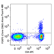
Alexa Fluor® 488 anti-human FOXP3

Human peripheral blood lymphocytes were surface stained with... 
-
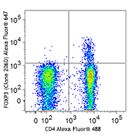
Alexa Fluor® 647 anti-human FOXP3

Human peripheral blood lymphocytes were surface stained with... 
-
FITC anti-human FOXP3

Human peripheral blood lymphocytes were surface stained with... 
-
Pacific Blue™ anti-human FOXP3

Human peripheral blood lymphocytes were surface stained with... 
-
PE anti-human FOXP3

Human peripheral blood lymphocytes were surface stained with... 
-
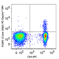
PE/Dazzle™ 594 anti-human FOXP3

Human peripheral blood lymphocytes were surface stained with... 
-
True-Nuclear™ One Step Staining Human Treg Flow™ Kit (FOXP3 Alexa Fluor® 488/CD25 PE/CD4 PerCP)

Human peripheral blood lymphocytes were stained with True-Nu... 
-
Brilliant Violet 421™ anti-human FOXP3

Human peripheral blood lymphocytes were surface stained with... 
-
KIRAVIA Blue 520™ anti-human FOXP3

Human peripheral blood lymphocytes were surface stained with... -
Spark NIR™ 685 anti-human FOXP3 Antibody

Human peripheral blood lymphocytes were surface stained with...


Follow Us