 Login / Register


- Clone
- EH12.2H7 (See other available formats)
- Regulatory Status
- RUO
- Other Names
- PD-1, PDCD1
- Isotype
- Mouse IgG1, κ
- Ave. Rating
- Submit a Review
- Product Citations
- publications

-

Human PBMCs were incubated for 3 days in media alone (top) or with PHA (bottom) and then stained with 170Er anti-CD3 (UCHT1) and 175Lu anti-CD279 (EH12.2H7). Data provided by DVS Sciences. -

| Cat # | Size | Price | Quantity Check Availability | Save | ||
|---|---|---|---|---|---|---|
| 329941 | 100 µg | 113€ | ||||
Programmed cell death 1 (PD-1), also known as CD279, is a 55 kD member of the immunoglobulin superfamily. CD279 contains the immunoreceptor tyrosine-based inhibitory motif (ITIM) in the cytoplasmic region and plays a key role in peripheral tolerance and autoimmune disease. CD279 is expressed predominantly on activated T cells, B cells, and myeloid cells. PD-L1 (B7-H1) and PD-L2 (B7-DC) are ligands of CD279 (PD-1) and are members of the B7 gene family. Evidence suggests overlapping functions for these two PD-1 ligands and their constitutive expression on some normal tissues and upregulation on activated antigen-presenting cells. Interaction of CD279 ligands results in inhibition of T cell proliferation and cytokine secretion.
Product DetailsProduct Details
- Verified Reactivity
- Human
- Reported Reactivity
- African Green, Baboon, Chimpanzee, Common Marmoset, Cynomolgus, Rhesus, Squirrel Monkey
- Antibody Type
- Monoclonal
- Host Species
- Mouse
- Formulation
- Phosphate-buffered solution, pH 7.2, containing 0.09% sodium azide and EDTA.
- Preparation
- The antibody was purified by affinity chromatography.
- Concentration
- 1.0 mg/ml
- Storage & Handling
- The antibody solution should be stored undiluted between 2°C and 8°C.
- Application
-
FC - Quality tested
CyTOF®, PG - Verified - Recommended Usage
-
This product is suitable for use with the Maxpar® Metal Labeling Kits. For metal labeling using Maxpar® Ready antibodies, proceed directly to the step to Partially Reduce the Antibody by adding 100 µl of Maxpar® Ready antibody to 100 µl of 4 mM TCEP-R in a 50 kDa filter and continue with the protocol. Always refer to the latest version of Maxpar® User Guide when conjugating Maxpar® Ready antibodies.
- Application Notes
-
Additional reported applications (for the relevant formats) include: blocking of ligand binding1-3, immunohistochemical staining of paraformaldehyde fixed frozen sections13, and spatial biology (IBEX)15,16. The LEAF™ purified antibody (Endotoxin <0.1 EU/µg, Azide-Free, 0.2 µm filtered) is recommended for functional assays (Cat. No. 329911 and 329912). For highly sensitive assays, we recommend Ultra-LEAF™ purified antibody (Cat. No. 329926) with a lower endotoxin limit than standard LEAF™ purified antibodies (Endotoxin <0.01 EU/µg).
- Additional Product Notes
-
Maxpar® is a registered trademark of Standard BioTools Inc.
- Application References
-
- Dorfman DM, et al. 2006 Am. J. Surg. Pathol. 30:802. (FA)
- Radziewicz H, et al. 2007. J. Virol. 81:2545. (FA)
- Velu V, et al. 2007. J. Virol. 81:5819. (FA)
- Zahn RC, et al. 2008. J. Virol. 82:11577. PubMed
- Chang WS, et al. 2008. J. Immunol. 181:6707. (FC) PubMed
- Nakamoto N, et al. 2009. PLoS Pathog. 5:e1000313. (FA)
- Jones RB, et al. 2009. J. Virol. 83:8722. (FC) PubMed
- Vojnov L, et al. 2010. J. Virol. 84:753. (FC) PubMed
- Radziewicz H, et al. 2010. J. Immunol. 184:2410. (FC) PubMed
- Monteriro P, et al. 2011. J. Immunol. 186:4618. PubMed
- Conrad J, et al. 2011. J. Immunol. 186:6871. PubMed
- Salisch NC, et al. 2010. J. Immunol. 184:476. (Rhesus reactivity)
- Li H and Pauza CD. 2015. Eur. J. Immunol. 45:298. (IHC)
- Peterson VM, et al. 2017. Nat. Biotechnol. 35:936. (PG)
- Radtke AJ, et al. 2020. Proc Natl Acad Sci USA. 117:33455-33465. (SB) PubMed
- Radtke AJ, et al. 2022. Nat Protoc. 17:378-401. (SB) PubMed
- Product Citations
-
- RRID
-
AB_2563734 (BioLegend Cat. No. 329941)
Antigen Details
- Structure
- Immunoglobulin superfamily
- Distribution
-
Transiently expressed on CD4- CD8- thymocytes; upregulated in thymocytes and splenic T and B lymphocytes; expressed on activated myeloid cells
- Ligand/Receptor
- B7-H1 (also known as PD-L1) and B7-DC (PD-L2)
- Cell Type
- B cells, Lymphocytes, T cells, Thymocytes, Tregs
- Biology Area
- Cancer Biomarkers, Immunology, Inhibitory Molecules
- Molecular Family
- CD Molecules, Immune Checkpoint Receptors
- Gene ID
- 5133 View all products for this Gene ID
- UniProt
- View information about CD279 on UniProt.org
Related Pages & Pathways
Pathways


Related FAQs
- Can I obtain CyTOF data related to your Maxpar® Ready antibody clones?
-
We do not test our antibodies by mass cytometry or on a CyTOF machine in-house. The data displayed on our website is provided by Fluidigm®. Please contact Fluidigm® directly for additional data and further details.
- Can I use Maxpar® Ready format clones for flow cytometry staining?
-
We have not tested the Maxpar® Ready antibodies formulated in solution containing EDTA for flow cytometry staining. While it is likely that this will work in majority of the situations, it is best to use the non-EDTA formulated version of the same clone for flow cytometry testing. The presence of EDTA in some situations might negatively affect staining.
- I am having difficulty observing a signal after conjugating a metal tag to your Maxpar® antibody. Please help troubleshoot.
-
We only supply the antibody and not test that in house. Please contact Fluidigm® directly for troubleshooting advice: http://techsupport.fluidigm.com/
- Is there a difference between buffer formulations related to Maxpar® Ready and purified format antibodies?
-
The Maxpar® Ready format antibody clones are formulated in Phosphate-buffered solution, pH 7.2, containing 0.09% sodium azide and EDTA. The regular purified format clones are formulated in solution that does not contain any EDTA. Both formulations are however without any extra carrier proteins.
Other Formats
View All CD279 Reagents Request Custom ConjugationCustomers Also Purchased
Compare Data Across All Formats
This data display is provided for general comparisons between formats.
Your actual data may vary due to variations in samples, target cells, instruments and their settings, staining conditions, and other factors.
If you need assistance with selecting the best format contact our expert technical support team.
-
Brilliant Violet 421™ anti-human CD279 (PD-1)

Human peripheral blood lymphocytes were stained with CD3 FIT... 
-
Purified anti-human CD279 (PD-1)

PHA-stimulated (day-3) human peripheral blood lymphocytes we... -
FITC anti-human CD279 (PD-1)

PHA-stimulated (day-3) human peripheral blood lymphocytes we... 
Human peripheral blood lymphocytes were stained with CD279 (... -
PE anti-human CD279 (PD-1)

PHA-stimulated (day-3) human peripheral blood lymphocytes we... 
Human peripheral blood lymphocytes were stained with CD279 (... 
Confocal image of human lymph node sample acquired using the... -
APC anti-human CD279 (PD-1)

PHA-stimulated (day-3) human peripheral blood lymphocytes we... 
Human peripheral blood lymphocytes were stained with CD279 (... -
Alexa Fluor® 647 anti-human CD279 (PD-1)

PHA-stimulated (day-3) human peripheral blood lymphocytes we... 
Human peripheral blood lymphocytes were stained with CD279 (... -
PerCP/Cyanine5.5 anti-human CD279 (PD-1)

PHA-stimulated (3-day) human peripheral blood lymphocytes we... 
Human peripheral blood lymphocytes were stained with CD3 APC... -
APC/Cyanine7 anti-human CD279 (PD-1)

PHA-stimulated (day-3) human peripheral blood lymphocytes st... -
Pacific Blue™ anti-human CD279 (PD-1)

Human peripheral blood lymphocytes were stained with CD279 (... 
PHA-stimulated (day-3) human peripheral blood lymphocytes we... -
PE/Cyanine7 anti-human CD279 (PD-1)

PHA-stimulated (day-3) human peripheral blood lymphocytes we... 
Human peripheral blood lymphocytes were stained with CD279 (... -
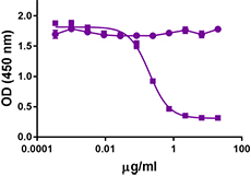
Purified anti-human CD279 (PD-1) (Maxpar® Ready)

Human PBMCs were incubated for 3 days in media alone (top) o... 
-
Brilliant Violet 605™ anti-human CD279 (PD-1)

Human peripheral blood lymphocytes were stained with CD3 FIT... -
Ultra-LEAF™ Purified anti-human CD279 (PD-1)
-
Brilliant Violet 711™ anti-human CD279 (PD-1)

Human peripheral blood lymphocytes were stained with CD3 FIT... -
Brilliant Violet 785™ anti-human CD279 (PD-1)

Human peripheral blood lymphocytes were stained with CD3 FIT... -
Brilliant Violet 510™ anti-human CD279 (PD-1)

PHA-stimulated (day-3) human peripheral blood lymphocytes we... -
Biotin anti-human CD279 (PD-1)

PHA-stimulated (3 days) human peripheral blood lymphocytes w... -
PE/Dazzle™ 594 anti-human CD279 (PD-1)

Human peripheral blood lymphocytes were stained with CD3 APC... 
PHA-stimulated (day 3) human peripheral blood lymphocytes st... 
-
Alexa Fluor® 488 anti-human CD279 (PD-1)

PHA-stimulated (day 3) human peripheral blood lymphocytes we... -
PerCP anti-human CD279 (PD-1)

PHA-stimulated (day 3) human peripheral blood lymphocytes we... -
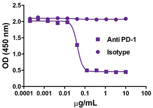
GoInVivo™ Purified anti-human CD279 (PD-1)

Anti-human PD-1 inhibits the binding of PD-L1. Immobilized P... -
Brilliant Violet 650™ anti-human CD279 (PD-1)

Human peripheral blood lymphocytes were stained with CD3 FIT... 
PHA-stimulated (three days) human peripheral blood lymphocyt... 
-
Alexa Fluor® 700 anti-human CD279 (PD-1)

Human peripheral blood lymphocytes were stained with CD3 FIT... 
PHA-stimulated (three days) human peripheral blood lymphocyt... 
-
APC/Fire™ 750 anti-human CD279 (PD-1)

PHA-stimulated (day 3) human peripheral blood lymphocytes we... -
TotalSeq™-A0088 anti-human CD279 (PD-1)
-
TotalSeq™-B0088 anti-human CD279 (PD-1)
-
TotalSeq™-C0088 anti-human CD279 (PD-1)
-
Brilliant Violet 750™ anti-human CD279 (PD-1)

PHA-stimulated (day 3) human peripheral blood lymphocytes we... -
TotalSeq™-D0088 anti-human CD279 (PD-1)
-
PE/Fire™ 640 anti-human CD279 (PD-1)

PHA stimulated (day 3) human peripheral blood lymphocytes we... 
Human peripheral blood lymphocytes were stained with anti-hu... -
PE/Cyanine5 anti-human CD279 (PD-1)

PHA-stimulated (3 days) human peripheral blood lymphocytes w...


Follow Us