 Login / Register


- Clone
- O93E9 (See other available formats)
- Regulatory Status
- RUO
- Other Names
- PYD and CARD domain-containing protein (PYCARD), Apoptosis-associated speck-like protein containing a CARD, Caspase recruitment domain protein 5, Target of methylation-induced silencing (TMS1)
- Isotype
- Mouse IgG1, κ
- Ave. Rating
- Submit a Review
- Product Citations
- publications

-

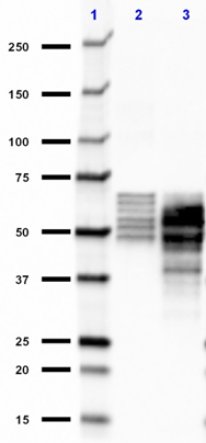
Total cell lysate of HL-60 (lane 1, 15 µg), HT-29 (lane 2, 15 µg), and mouse thymus (lane 3, 15 µg) were resolved by electrophoresis, transferred to nitrocellulose, and probed with purified monoclonal anti-ASC (clone O93E9) antibody. Proteins were visualized using a goat anti-mouse-IgG secondary antibody conjugated to HRP and chemiluminescence detection.
| Cat # | Size | Price | Quantity Check Availability | Save | ||
|---|---|---|---|---|---|---|
| 676502 | 100 µg | 249€ | ||||
Apoptosis-associated speck-like protein containing a CARD (ASC), also known as target of methylation-mediated silencing (TMS1), PYD, and CARD domain containing protein (PYCARD), is a 21.6 kD pro-apoptotic protein that contains a N-terminal pyrin domain (PYD) and a C-terminal caspase recruitment domain (CARD). The TMS1 gene was originally found to be aberrantly methylated and silenced in various cancer cells. Expression of ASC can be induced by pro-apoptotic/inflammatory stimuli. In normal cells, this protein is localized to the cytoplasm. However, in cells undergoing apoptosis, it forms ball-like aggregates near the nuclear periphery. During apoptosis, ASC translocates from the cytosol to the mitochondria and associates with mitochondrial Bax to trigger cytochrome c release and subsequent apoptosis. The adaptor molecule ASC mediates AIM2-dependent caspase-1 activation. Also, the AIM2/ASC complex can act as a novel caspase-8 activation platform. Recent studes has shown that ASC reduces the phosphorylation of IKKα/β phosphorylation and inhibits NF-κB activity in primary melanoma. However, in metastatic melanoma through, it has the opposite function enhancing NF-κB activity and the secretion of IL-1β.
Product DetailsProduct Details
- Verified Reactivity
- Human
- Antibody Type
- Monoclonal
- Host Species
- Mouse
- Immunogen
- Full length recombinant human PYCARD (NP_037390) produced in E. coli.
- Formulation
- Phosphate-buffered solution, pH 7.2, containing 0.09% sodium azide.
- Preparation
- The antibody was purified by affinity chromatography.
- Concentration
- 0.5 mg/ml
- Storage & Handling
- The antibody solution should be stored undiluted between 2°C and 8°C.
- Application
-
WB - Quality tested
- Recommended Usage
-
Each lot of this antibody is quality control tested by Western blotting. For Western blotting, the suggested use of this reagent is 0.5 - 2.0 µg per ml. It is recommended that the reagent be titrated for optimal performance for each application.
- Product Citations
-
- RRID
-
AB_2565643 (BioLegend Cat. No. 676502)
Antigen Details
- Structure
- Full-length ASC (fASC) contains 195 amino acids wtih a predicted molecular weight of 21.6 kD. The sequence of isoform ASC-b is missing 93-111 a.a. The sequence of isoform ASC-c is missing 26-85 a.a.
- Distribution
-
Cytoplasm. In resting monocytes and macrophages, the nucleus is the primary localization.
- Function
- ASC promotes caspase-mediated apoptosis. This pro-apoptotic activity is mediated predominantly through the activation of caspase-9. ASC is an integral component of the inflammasome. Within the inflammasome, ASC links caspase-1 and NOD-like receptors (NLR), which lead to the activation of caspase-1.
- Ligand/Receptor
- CIAS1/PYPAF1 and PYDC1.
- Cell Type
- Dendritic cells
- Biology Area
- Angiogenesis, Cell Biology, Immunology, Innate Immunity, Neuroscience, Signal Transduction
- Antigen References
-
1. Masumoto J, et al. 1999. J. Biol. Chem. 274:33835.
2. Conway KE, et al. 2000. Cancer Res. 60:6236.
3. Strong R, et al. 1991. Brain Res. 542:23.
4. Srinivasula SM, et al. 2002. J. Biol. Chem. 277:21119.
5. Ohtsuka T, et al. 2004. Nat. Cell Biol. 6:121.
6. Ippagunta SK, et al. 2010. J. Biol. Chem. 285:12454.
7. Liu W, et al. 2013. J. Invest Dermatol. 2:518-527. - Gene ID
- 29108 View all products for this Gene ID
- UniProt
- View information about ASC on UniProt.org

Related FAQs
Other Formats
View All ASC Reagents Request Custom Conjugation| Description | Clone | Applications |
|---|---|---|
| Purified anti-ASC | O93E9 | WB |
Customers Also Purchased
Compare Data Across All Formats
This data display is provided for general comparisons between formats.
Your actual data may vary due to variations in samples, target cells, instruments and their settings, staining conditions, and other factors.
If you need assistance with selecting the best format contact our expert technical support team.
-
Purified anti-ASC

Total cell lysate of HL-60 (lane 1, 15 µg), HT-29 (lane 2, 1...


Follow Us