 Login / Register


- Clone
- OX-7 (See other available formats)
- Regulatory Status
- RUO
- Other Names
- Thy-1, Thy-1.1
- Isotype
- Mouse IgG1, κ
- Ave. Rating
- Submit a Review
- Product Citations
- publications

-

LOU rat thymocytes stained with OX-7 PE
| Cat # | Size | Price | Quantity Check Availability | Save | ||
|---|---|---|---|---|---|---|
| 202523 | 50 µg | 59€ | ||||
| 202524 | 200 µg | 144€ | ||||
CD90, also known as Thy-1, is a 28-30 kD GPI-linked membrane glycoprotein. It is a member of the immunoglobulin superfamily and has been shown to interact with CD45 in signal transduction during lymphocyte proliferation and differentiation. CD90 is expressed on hematopoietic stem cells, neurons, thymocytes, peripheral T cells, fibroblasts, stromal cells.
Product DetailsProduct Details
- Verified Reactivity
- Rat, Mouse
- Reported Reactivity
- Rabbit, Guinea Pig
- Antibody Type
- Monoclonal
- Host Species
- Mouse
- Immunogen
- Rat thymocyte Thy-1 antigen
- Formulation
- Phosphate-buffered solution, pH 7.2, containing 0.09% sodium azide.
- Preparation
- The antibody was purified by affinity chromatography, and conjugated with PE under optimal conditions.
- Concentration
- 0.2 mg/ml
- Storage & Handling
- The antibody solution should be stored undiluted between 2°C and 8°C, and protected from prolonged exposure to light. Do not freeze.
- Application
-
FC - Quality tested
- Recommended Usage
-
Each lot of this antibody is quality control tested by immunofluorescent staining with flow cytometric analysis. For flow cytometric staining, the suggested use of this reagent is ≤0.25 µg per million cells in 100 µl volume. It is recommended that the reagent be titrated for optimal performance for each application.
- Excitation Laser
-
Blue Laser (488 nm)
Green Laser (532 nm)/Yellow-Green Laser (561 nm)
- Application Notes
-
The OX-7 antibody reacts with rat CD90 and mouse CD90.1 (Thy-1.1) (which is expressed by mouse strains of AKR/J, PL, and FVB/N), but not mouse CD90.2.
Additional reported applications (for the relevant formats) include: immunohistochemical7 and immunofluorescent8 staining of acetone-fixed frozen sections and zinc-fixed paraffin-embedded sections, immunoprecipitation1, Western blotting1, in vitro activation of leukocytes2, induction of endothelial cell permeability3, induction of apoptosis in glomerular mesangial cells, and induction of glomerulonephritis in vivo4. - Application References
-
- Jeng CJ, et al. 1998. J. Cell Biol. 140:685. (IP, WB)
- Nakashima I, et al. 1991. J. Immunol. 147:1153.
- Ishizu A, et al. 1995. Int. Immunol. 7:1939.
- Eitner F. 1997. Kidney. Int. 51:69.
- Kawachi H, et al. 1992. Clin. Exp. Immunol. 88:399. (WB)
- Dyer KD, et al. 2007. J. Immunol. 179:1693. (FC) PubMed
- Daniel C, et al. 2012. Lab Invest. 92:812. (IHC-P)
- Li B, et al. 2006. Kidney Int. 69:323. (ICC)
- Uchimura H, et al. 2005. J Am Soc Nephrol. 16(4):997-1004. (IHC-F)
- Inagi R, et al. 2008. J Am Soc Nephrol. 19(5):915-22. (IHC-P)
- Product Citations
-
- RRID
-
AB_1595635 (BioLegend Cat. No. 202523)
AB_1595524 (BioLegend Cat. No. 202524)
Antigen Details
- Structure
- Ig superfamily, GPI-anchored membrane glycoprotein, 28-30 kD
- Distribution
-
Hematopoietic stem cells, thymocytes, a subset of peripheral blood T cells, early myeloid and erythroid progenitors, immature B cells, neurons, activated endothelium, mast cells, dendritic cells, glomeruli
- Function
- Adhesion, signal transduction, lymphocyte co-stimulation, proliferation and differentiation of hematopoietic stem cells
- Ligand/Receptor
- Interacts with CD45
- Cell Type
- B cells, Dendritic cells, Endothelial cells, Hematopoietic stem and progenitors, Mast cells, Neurons, T cells, Thymocytes
- Biology Area
- Immunology
- Molecular Family
- CD Molecules
- Antigen References
-
1. Campbell DG, et al. 1981. Biochem. J. 195:15.
2. Hosseinzadeh H, et al. 1993. J. Immunol. 150:1670. - Gene ID
- 21838 View all products for this Gene ID 24832 View all products for this Gene ID
- UniProt
- View information about CD90/CD90.1 on UniProt.org


Related FAQs
- What type of PE do you use in your conjugates?
- We use R-PE in our conjugates.
Other Formats
View All CD90/CD90.1 Reagents Request Custom ConjugationCustomers Also Purchased
Compare Data Across All Formats
This data display is provided for general comparisons between formats.
Your actual data may vary due to variations in samples, target cells, instruments and their settings, staining conditions, and other factors.
If you need assistance with selecting the best format contact our expert technical support team.
-
PE/Cyanine7 anti-rat CD90/mouse CD90.1 (Thy-1.1)

LOU rat thymocytes stained with OX-7 PE/Cyanine7 -
Purified anti-rat CD90/mouse CD90.1 (Thy-1.1)

LOU rat thymocytes stained with purified OX-7, followed by a... 
FVB/N mouse frozen thymus section was fixed with 4% paraform... -
FITC anti-rat CD90/mouse CD90.1 (Thy-1.1)

Lou rat thymocytes stained with OX-7 FITC -
Alexa Fluor® 488 anti-rat CD90/mouse CD90.1 (Thy-1.1)

Rat thymocytes stained with OX-7 Alexa Fluor® 488 
FVB/N mouse frozen thymus section was fixed with 4% paraform... -
Alexa Fluor® 647 anti-rat CD90/mouse CD90.1 (Thy-1.1)

LOU rat thymocytes stained with OX-7 Alexa Fluor® 647 
FVB/N mouse frozen thymus section was fixed with 4% paraform... -
PerCP/Cyanine5.5 anti-rat CD90/mouse CD90.1 (Thy-1.1)

Lewis rat thymocytes were stained with CD3 APC and CD90/mous... -
APC/Cyanine7 anti-rat CD90/mouse CD90.1 (Thy-1.1)

LOU rat splenocytes stained with OX-7 APC/Cyanine7 
LOU rat thymocytes stained with OX-7 APC/Cyanine7 -
Pacific Blue™ anti-rat CD90/mouse CD90.1 (Thy-1.1)

LOU rat thymocytes stained with OX-7 Pacific Blue™ -
PerCP anti-rat CD90/mouse CD90.1 (Thy-1.1)

Lou rat thymocytes stained with OX-7 PerCP -
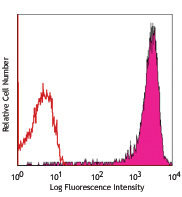
PE anti-rat CD90/mouse CD90.1 (Thy-1.1)

LOU rat thymocytes stained with OX-7 PE -
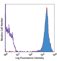
APC anti-rat CD90/mouse CD90.1 (Thy-1.1)

Lou rat thymocytes stained with OX-7 APC -
Alexa Fluor® 700 anti-rat CD90/mouse CD90.1 (Thy-1.1)

LOU rat thymocytes stained with OX-7 Alexa Fluor® 700 -
Biotin anti-rat CD90/mouse CD90.1 (Thy-1.1)

LOU rat thymocytes stained with OX-7 biotin, followed by Sav... -
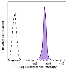
Brilliant Violet 421™ anti-rat CD90/mouse CD90.1 (Thy-1.1)

FVB mouse splenocytes were stained with CD90.1 (clone OX-7) ... 
C57BL/6 mouse splenocytes were stained with CD90.1 (clone OX... -
Brilliant Violet 650™ anti-rat CD90/mouse CD90.1 (Thy-1.1)

Lou rat thymocytes were stained with CD90.1 (clone OX-7) Bri... -
Brilliant Violet 510™ anti-rat CD90/mouse CD90.1 (Thy-1.1)

FVB mouse splenocytes were stained with CD90.1 (clone OX-7) ... 
Lewis rat thymocytes were stained with CD90.1 (clone OX-7) B... -
Brilliant Violet 605™ anti-rat CD90/mouse CD90.1 (Thy-1.1)

FVB/N mouse splenocytes were stained with CD90.1 (clone OX-7... 
Lewis rat thymocytes were stained with CD90.1 (clone OX-7) B... -
Brilliant Violet 711™ anti-rat CD90/mouse CD90.1 (Thy-1.1)

FVB/N mouse splenocytes were stained with CD90.1 (clone OX-7... 
Lewis rat thymocytes were stained with CD90.1 (clone OX-7) B... -
PE/Dazzle™ 594 anti-rat CD90/mouse CD90.1 (Thy-1.1)

FVB mouse splenocytes were stained with CD90.1 (clone OX-7) ... -
APC/Fire™ 750 anti-rat CD90/mouse CD90.1 (Thy-1.1)

Lewis rat thymocytes were stained with APC/Fire™ 750 anti-ra... -
Alexa Fluor® 594 anti-rat CD90/mouse CD90.1 (Thy-1.1)

FVB/N mouse frozen thymus section was fixed with 4% paraform... -
TotalSeq™-A0380 anti-rat CD90/mouse CD90.1 (Thy-1.1)
-
TotalSeq™-B0380 anti-rat CD90/mouse CD90.1 (Thy-1.1)
-
TotalSeq™-C0380 anti-rat CD90/mouse CD90.1 (Thy-1.1)
-
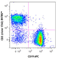
Brilliant Violet 785™ anti-rat CD90/mouse CD90.1 (Thy-1.1)

Lewis rat thymocytes were stained with anti-rat CD3 FITC and...


Follow Us