 Login / Register


- Clone
- ASC-1 (See other available formats)
- Regulatory Status
- RUO
- Workshop
- VI A002
- Other Names
- α3 integrin, integrin α3 chain, VLA-3 α chain, VLA3a, ITGA3
- Isotype
- Mouse IgG1, κ
- Ave. Rating
- Submit a Review
- Product Citations
- publications

-

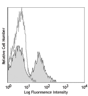
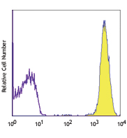
Human cervical cancer cell line HeLa cells stained with ASC-1 PE
| Cat # | Size | Price | Quantity Check Availability | Save | ||
|---|---|---|---|---|---|---|
| 343803 | 25 tests | 117€ | ||||
CD49c is a 150 kD α integrin chain known as α3 integrin or VLA-3 α chain. It is a type I transmembrane glycoprotein which is proteolytically cleaved into two disulfide linked fragments of 125 kD and 30 kD. CD49c forms a heterodimer with integrin β1 (α3β1, CD49c/CD29, VLA-3) and is expressed by many types of adhesion cells, such as endothelial cells, epithelial cells, and dermal fibroblasts. Weak expression has been reported on leukocytes. VLA-3 plays a role in cell-cell and cell-matrix adhesion through binding Kalinin, collagen, laminin-1, laminin-5, entactin, and fibronectin.
Product DetailsProduct Details
- Verified Reactivity
- Human
- Antibody Type
- Monoclonal
- Host Species
- Mouse
- Immunogen
- Human squamous cell carcinoma cell line SCC-9
- Formulation
- Phosphate-buffered solution, pH 7.2, containing 0.09% sodium azide and BSA (origin USA)
- Preparation
- The antibody was purified by affinity chromatography, and conjugated with PE under optimal conditions.
- Concentration
- Lot-specific (to obtain lot-specific concentration, please enter the lot number in our Concentration and Expiration Lookup or Certificate of Analysis online tools.)
- Storage & Handling
- The antibody solution should be stored undiluted between 2°C and 8°C, and protected from prolonged exposure to light. Do not freeze.
- Application
-
FC - Quality tested
- Recommended Usage
-
Each lot of this antibody is quality control tested by immunofluorescent staining with flow cytometric analysis. For flow cytometric staining, the suggested use of this reagent is 5 µl per million cells in 100 µl staining volume or 5 µl per 100 µl of whole blood.
- Excitation Laser
-
Blue Laser (488 nm)
Green Laser (532 nm)/Yellow-Green Laser (561 nm)
- Application Notes
-
Additional reported applications include: immunoprecipitation4, Western blotting5, and immunohistochemical staining of frozen tissue sections2.
- Application References
-
- Pattaramalai S, et al. 1996. Exp. Cell. Res. 222:281.
- Skubitz AP, et al. 1996. Am. J. Pathol. 148:1445. (IHC)
- Skubitz AP, et al. 1998. FEBS Lett. 426:386.
- Calzada MJ, et al. 2004. Circ. Res. 94:462. (IP)
- Zhang X, et al. 2003. Surgery. 133:429. (WB)
- Product Citations
-
- RRID
-
AB_1731941 (BioLegend Cat. No. 343803)
Antigen Details
- Structure
- 150 kD type I transmembrane glycoprotein, integrin family, associate with integrin β1(CD29)
- Distribution
- Endothelial cells, epithelial cells, dermal fibroblast
- Function
- adhesion
- Ligand/Receptor
- Kalinin, collagen, laminin-1, laminin-5 and entactin, fibronectin
- Cell Type
- Endothelial cells, Epithelial cells, Fibroblasts
- Biology Area
- Cell Biology, Immunology, Neuroscience, Synaptic Biology
- Molecular Family
- Adhesion Molecules, CD Molecules
- Antigen References
-
1. Zola H, et al. 2007. Leukocyte and stromal Cell Molecules:the CD Markers. John Wiley & Sons Inc.
- Gene ID
- 3675 View all products for this Gene ID
- UniProt
- View information about CD49c on UniProt.org


Related FAQs
- What type of PE do you use in your conjugates?
- We use R-PE in our conjugates.
Other Formats
View All CD49c Reagents Request Custom Conjugation| Description | Clone | Applications |
|---|---|---|
| Purified anti-human CD49c (integrin α3) | ASC-1 | FC,IP,WB,IHC-F |
| PE anti-human CD49c (integrin α3) | ASC-1 | FC |
| APC anti-human CD49c (integrin α3) | ASC-1 | FC |
Customers Also Purchased

Compare Data Across All Formats
This data display is provided for general comparisons between formats.
Your actual data may vary due to variations in samples, target cells, instruments and their settings, staining conditions, and other factors.
If you need assistance with selecting the best format contact our expert technical support team.
-
Purified anti-human CD49c (integrin α3)

Human cervical cancer cell line HeLa cells stained with puri... -
PE anti-human CD49c (integrin α3)
Human cervical cancer cell line HeLa cells stained with ASC-... -
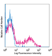
APC anti-human CD49c (integrin α3)

Human cervical cancer cell line HeLa cells stained with ASC-...


Follow Us