 Login / Register


- Clone
- 2E7 (See other available formats)
- Regulatory Status
- RUO
- Other Names
- Integrin αIEL chain, Integrin αE chain, αE integrin, ITGAE
- Isotype
- Armenian Hamster IgG
- Ave. Rating
- Submit a Review
- Product Citations
- publications

-

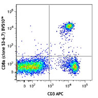
C57BL/6 mouse splenocytes were stained with CD3 APC and CD103 (clone 2E7) Brilliant Violet 510™ (top) or Armenian hamster IgG Brilliant Violet 510™ isotype control (bottom). -

| Cat # | Size | Price | Quantity Check Availability | Save | ||
|---|---|---|---|---|---|---|
| 121423 | 50 µg | 229€ | ||||
CD103 is a type I transmembrane glycoprotein known as αE integrin or Integrin αIEL chain. It belongs to the integrin family and is primarily found on intestinal intraepithelial lymphocytes (IEL). CD103 is also expressed on a subpopulation of lamina propria T cells, epithelial dendritic cells, lamina propria-derived dendritic cells, and a small subset of peripheral lymphocytes. T regulatory cells express high level of CD103. The CD103 expression on lymphocytes can be induced upon activation and TGF-β stimulation. In association with integrin β7, CD103 is expressed as αE/β7 heterodimer. Mature CD103 protein can be cleaved into 2 chains, a 150 kD (C-terminal) chain and a 25 kD (N-terminal) chain, which remain linked by disulfide bonds. CD103 binds to E-cadherin and mediates homing of lymphocytes to the intestinal epithelium.
Product DetailsProduct Details
- Verified Reactivity
- Mouse
- Antibody Type
- Monoclonal
- Host Species
- Armenian Hamster
- Immunogen
- Mouse intestinal intraepithelial lymphocytes
- Formulation
- Phosphate-buffered solution, pH 7.2, containing 0.09% sodium azide and BSA (origin USA).
- Preparation
- The antibody was purified by affinity chromatography and conjugated with Brilliant Violet 510™ under optimal conditions.
- Concentration
- 0.2 mg/ml
- Storage & Handling
- The antibody solution should be stored undiluted between 2°C and 8°C, and protected from prolonged exposure to light. Do not freeze.
- Application
-
FC - Quality tested
- Recommended Usage
-
Each lot of this antibody is quality control tested by immunofluorescent staining with flow cytometric analysis. For flow cytometric staining, the suggested use of this reagent is ≤0.5 µg per million cells in 100 µl volume. It is recommended that the reagent be titrated for optimal performance for each application.
Brilliant Violet 510™ excites at 405 nm and emits at 510 nm. The bandpass filter 510/50 nm is recommended for detection, although filter optimization may be required depending on other fluorophores used. Be sure to verify that your cytometer configuration and software setup are appropriate for detecting this channel. Refer to your instrument manual or manufacturer for support. Brilliant Violet 510™ is a trademark of Sirigen Group Ltd.
Learn more about Brilliant Violet™.
This product is subject to proprietary rights of Sirigen Inc. and is made and sold under license from Sirigen Inc. The purchase of this product conveys to the buyer a non-transferable right to use the purchased product for research purposes only. This product may not be resold or incorporated in any manner into another product for resale. Any use for therapeutics or diagnostics is strictly prohibited. This product is covered by U.S. Patent(s), pending patent applications and foreign equivalents. - Excitation Laser
-
Violet Laser (405 nm)
- Application Notes
-
Additional reported applications (for the relevant formats) include: immunoprecipitation1, immunohistochemical staining1,7 of acetone-fixed frozen sections, immunofluorescence2, and in vitro activation1.
- Application References
-
- LeFrancois L, et. al, 1994. Eur. J. Immunol. 24:635. (FC, IHC, IP)
- Mysorekar IU, et. al, 2002. J. Biol. Chem. 277:37811. (FC, IF)
- Mikami N, et al. 2011. J. Immunol. 186:6886. PubMed
- del Rio ML, et al. 2011. Transpl. Int. 24:501. (FC) PubMed
- Quinn KM, et al. 2013. J. Immunol. 191:5085. PubMed
- Verhagen J and Wraith DC. 2014. J. Immunol. Methods. S0022. (FC) PubMed
- Xiao B, et al. 2015. PLoS One 1:e0115333. (IHC)
- Product Citations
-
- RRID
-
AB_2562713 (BioLegend Cat. No. 121423)
Antigen Details
- Structure
- Type I transmembrane glycoprotein, Integrin family, can be cleaved into 150 kD and 25 kD chains, associated with β7 integrin
- Distribution
-
Majority of intestinal intraepithelial lymphocytes (IEL), subpopulation of lamina propria T cells, epithelial dendritic cells, small subset of peripheral lymphocytes, Treg cells.
- Function
- Retention and activation of CD103+ lymphocytes in the intestinal epithelium, regulate tissue-specific T cell homing.
- Ligand/Receptor
- E-Cadherin
- Cell Type
- Dendritic cells, Lymphocytes, T cells, Tregs
- Biology Area
- Immunology
- Molecular Family
- Adhesion Molecules, CD Molecules
- Antigen References
-
1. Kilshaw PJ and SJ. Murant. 1990. Eur. J. Immunol. 20:2201.
2. Karecla PI, et al. 1995. Eur. J. Immunol. 25:852.
3. LeFrancois L, et al. 1994. Eur. J. Immunol. 24:635.
4. Sung SS, et al. 2006. J. Immunol. 176:2161.
5. Johansson-Lindbom B, et al. 2005. J. Exp. Med. 202:1063.
6. Dujardin HC, et al. 2004. Proc. Natl. Acad. Sci. USA. 101:14473. - Gene ID
- 16407 View all products for this Gene ID
- UniProt
- View information about CD103 on UniProt.org
Related Pages & Pathways
Pathways


Related FAQs
Other Formats
View All CD103 Reagents Request Custom Conjugation| Description | Clone | Applications |
|---|---|---|
| Purified anti-mouse CD103 | 2E7 | FC,IHC-F,IP |
| Biotin anti-mouse CD103 | 2E7 | FC |
| PE anti-mouse CD103 | 2E7 | FC |
| Alexa Fluor® 488 anti-mouse CD103 | 2E7 | FC |
| Alexa Fluor® 647 anti-mouse CD103 | 2E7 | FC |
| Alexa Fluor® 594 anti-mouse CD103 | 2E7 | IHC-F |
| Brilliant Violet 510™ anti-mouse CD103 | 2E7 | FC |
| APC anti-mouse CD103 | 2E7 | FC |
| PerCP/Cyanine5.5 anti-mouse CD103 | 2E7 | FC |
| Pacific Blue™ anti-mouse CD103 | 2E7 | FC |
| FITC anti-mouse CD103 | 2E7 | FC |
| Brilliant Violet 421™ anti-mouse CD103 | 2E7 | FC |
| PE/Cyanine7 anti-mouse CD103 | 2E7 | FC |
| APC/Cyanine7 anti-mouse CD103 | 2E7 | FC |
| PE/Dazzle™ 594 anti-mouse CD103 | 2E7 | FC |
| Brilliant Violet 605™ anti-mouse CD103 | 2E7 | FC |
| Brilliant Violet 711™ anti-mouse CD103 | 2E7 | FC |
| TotalSeq™-A0201 anti-mouse CD103 | 2E7 | PG |
| Brilliant Violet 785™ anti-mouse CD103 | 2E7 | FC |
| Alexa Fluor® 700 anti-mouse CD103 | 2E7 | FC |
| TotalSeq™-C0201 anti-mouse CD103 | 2E7 | PG |
| TotalSeq™-B0201 anti-mouse CD103 | 2E7 | PG |
| PE/Cyanine5 anti-mouse CD103 | 2E7 | FC |
| Spark Red™ 718 anti-mouse CD103 (Flexi-Fluor™) | 2E7 | FC |
Customers Also Purchased
Compare Data Across All Formats
This data display is provided for general comparisons between formats.
Your actual data may vary due to variations in samples, target cells, instruments and their settings, staining conditions, and other factors.
If you need assistance with selecting the best format contact our expert technical support team.
-
Purified anti-mouse CD103

C57BL/6 mouse splenocytes stained with PE CD3 (clone 17A2) a... -
Biotin anti-mouse CD103

C57BL/6 mouse splenocytes stained with CD3 (145-2C11) FITC a... -
PE anti-mouse CD103

C57BL/6 splenocytes stained with 2E7 PE -
Alexa Fluor® 488 anti-mouse CD103

C57BL/6 mouse splenocytes stained with PE CD3 (clone 17A2) a... -
Alexa Fluor® 647 anti-mouse CD103

C57BL/6 mouse splenocytes stained with PE CD3 (clone 17A2) a... -
Alexa Fluor® 594 anti-mouse CD103

C57BL/6 mouse frozen intestine section was fixed with cold a... -
Brilliant Violet 510™ anti-mouse CD103

C57BL/6 mouse splenocytes were stained with CD3 APC and CD10... 
-
APC anti-mouse CD103

BALB/c splenocytes stained with 2E7 APC -
PerCP/Cyanine5.5 anti-mouse CD103

C57BL/6 mouse splenocytes were stained with CD3 APC and CD10... -
Pacific Blue™ anti-mouse CD103

C57BL/6 splenocytes stained with 2E7 Pacific Blue™ and... 
C57BL/6 splenocytes stained with Armenian Hamster isotype co... -
FITC anti-mouse CD103

C57BL/6 mouse splenocytes were stained with CD3 APC and CD10... 
-
Brilliant Violet 421™ anti-mouse CD103

C57BL/6 mouse splenocytes were stained with CD3 FITC and CD1... 
-
PE/Cyanine7 anti-mouse CD103

C57BL/6 mouse splenocytes were stained with CD3 APC and CD10... -
APC/Cyanine7 anti-mouse CD103

C57BL/6 mouse splenocytes were stained with CD3 PE and CD103... -
PE/Dazzle™ 594 anti-mouse CD103

C57BL/6 mouse splenocytes were stained with CD3 APC and CD10... 
-
Brilliant Violet 605™ anti-mouse CD103

C57BL/6 mouse splenocytes were stained with CD3 APC and CD10... 
-
Brilliant Violet 711™ anti-mouse CD103

C57BL/6 mouse splenocytes were stained with CD3 PE and CD103... 
-
TotalSeq™-A0201 anti-mouse CD103
-
Brilliant Violet 785™ anti-mouse CD103

C57BL/6 mouse splenocytes were stained with CD3 FITC and CD1... -
Alexa Fluor® 700 anti-mouse CD103

C57BL/6 mouse splenocytes were stained with CD3 PE and CD103... -
TotalSeq™-C0201 anti-mouse CD103
-
TotalSeq™-B0201 anti-mouse CD103
-
PE/Cyanine5 anti-mouse CD103

C57BL/6 mouse splenocytes were stained with anti-mouse CD3 F... -
Spark Red™ 718 anti-mouse CD103 (Flexi-Fluor™)


Follow Us