 Login / Register


- Clone
- S16007D (See other available formats)
- Regulatory Status
- RUO
- Other Names
- P2Y12, ADPG-R, BDPLT8, HORK3, P2T(AC), P2Y(12)R, P2Y(AC), P2Y(ADP), P2Y(cyc), P2Y12P2Y12 platelet ADP receptor, purinergic receptor P2RY12, purinergic receptor P2Y, G-protein coupled, 12
- Isotype
- Rat IgG2b, κ
- Ave. Rating
- Submit a Review
- Product Citations
- publications

-

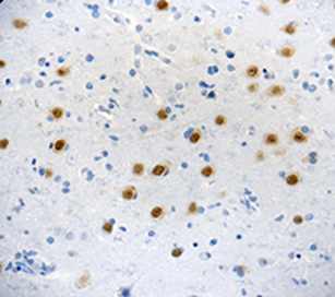
IHC staining of Biotin anti-P2RY12 antibody (clone S16007D) on formalin-fixed paraffin-embedded mouse brain tissue. Following antigen retrieval using Retrieve‐All Antigen Unmasking System 3 (Cat. No. 927801), the tissue was incubated with the primary antibody at 10 µg/mL overnight at 4°C, followed by incubation with 2.5 µg/ml of Alexa Fluor® 647 Streptavidin (Cat. No. 405237) for 1 hour at room temperature. Nuclei were counterstained with DAPI. The image was captured with a 40X objective. Scale bar: 50 µm -

IHC staining of Biotin anti-P2RY12 antibody (clone S16007D) on formalin-fixed paraffin-embedded mouse hippocampal tissue. Following antigen retrieval using Retrieve‐All Antigen Unmasking System 3 (Cat. No. 927801), the tissue was incubated with the primary antibody at 10 µg/mL overnight at 4°C, followed by incubation with 2.5 µg/ml of Alexa Fluor® 647 Streptavidin (Cat. No. 405237) for 1 hour at room temperature. Nuclei were counterstained with DAPI. The image was captured with a 10X objective. Scale bar: 100 µm
| Cat # | Size | Price | Quantity Check Availability | Save | ||
|---|---|---|---|---|---|---|
| 848008 | 25 µg | 109€ | ||||
| 848007 | 100 µg | 254€ | ||||
Purinergic Receptor P2Y (P2RY12) is a receptor for ADP and ATP coupled to G-proteins that inhibit the adenylyl cyclase second messenger system. P2RY12 is not activated by UDP and UTP. Required for normal platelet aggregation and blood coagulation. P2RY12 is a highly selective marker for microglia that specifically distinguishes these cells from other myeloid cells.
Product DetailsProduct Details
- Verified Reactivity
- Mouse
- Antibody Type
- Monoclonal
- Host Species
- Rat
- Immunogen
- Mouse P2RY12.
- Formulation
- Phosphate-buffered solution, pH 7.2, containing 0.09% sodium azide.
- Preparation
- The antibody was purified by affinity chromatography and conjugated with biotin under optimal conditions.
- Concentration
- 0.5 mg/ml
- Storage & Handling
- The antibody solution should be stored undiluted between 2°C and 8°C. Do not freeze.
- Application
-
IHC-P - Quality tested
- Recommended Usage
-
Each lot of this antibody is quality control tested by formalin-fixed paraffin-embedded immunohistochemical staining. For immunohistochemistry, a concentration range of 5.0 - 10 µg/ml is suggested. It is recommended that the reagent be titrated for optimal performance for each application.
- Application Notes
-
View the video of our collaborator discussing the importance of highly specific monoclonal antibodies for P2RY12 in Microglia research.
Learn more about the validation testing we performed on clone S16007D by watching our Scientific Poster Video. - RRID
-
AB_2749911 (BioLegend Cat. No. 848008)
AB_2749906 (BioLegend Cat. No. 848007)
Antigen Details
- Structure
- P2RY12 is a 347 amino acid protein with a molecular mass of 39.8 kD.
- Distribution
-
Tissue distribution: platelets, brain (microglia), lung, appendix, pituitary and adrenal glands.
Cellular distribution: cell membrane and cytosol. - Function
- G-protein coupled receptor that inhibits the adenylyl cyclase second messenger system.
- Ligand/Receptor
- ADP and ATP.
- Cell Type
- Microglia, Platelets
- Biology Area
- Cell Biology, Immunology, Innate Immunity, Neuroscience, Neuroscience Cell Markers
- Molecular Family
- GPCR, Purinergic Receptors
- Antigen References
-
1. Orr AG, et al. 2009. Nat. Neurosci. 12(7):872-8.
- Gene ID
- 70839 View all products for this Gene ID
- UniProt
- View information about P2RY12 on UniProt.org


Related FAQs
- How many biotin molecules are per antibody structure?
- We don't routinely measure the number of biotins with our antibody products but the number of biotin molecules range from 3-6 molecules per antibody.
Other Formats
View All P2RY12 Reagents Request Custom Conjugation| Description | Clone | Applications |
|---|---|---|
| Purified anti-P2RY12 | S16007D | IHC-P,FC |
| PE anti-P2RY12 | S16007D | FC |
| APC anti-P2RY12 | S16007D | FC |
| Biotin anti-P2RY12 | S16007D | IHC-P |
| TotalSeq™-A0415 anti-P2RY12 | S16007D | PG |
| Spark YG™ 570 anti-P2RY12 Antibody | S16007D | IHC-P |
| APC/Fire™ 810 anti-P2RY12 | S16007D | FC |
| Alexa Fluor® 488 anti-P2RY12 | S16007D | FC |
| Alexa Fluor® 647 anti-P2RY12 | S16007D | FC |
| TotalSeq™-B0415 anti-P2RY12 | S16007D | PG |
| TotalSeq™-C0415 anti-P2RY12 | S16007D | PG |
Customers Also Purchased
Compare Data Across All Formats
This data display is provided for general comparisons between formats.
Your actual data may vary due to variations in samples, target cells, instruments and their settings, staining conditions, and other factors.
If you need assistance with selecting the best format contact our expert technical support team.
-
Purified anti-P2RY12

IHC staining of purified anti-P2RY12 antibody (clone S16007D... 
IHC staining of purified anti-P2RY12 antibody (clone S16007D... 
IHC staining of purified anti-P2RY12 antibody (clone S16007D... 
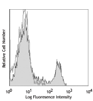
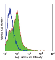
Single cell suspension of mouse brain tissue was stained wit... -
PE anti-P2RY12

Brain from a C57BL/6 mouse was digested with collagenase and... 
-
APC anti-P2RY12

Brain from a C57BL/6 mouse was digested with collagenase and... 
-
Biotin anti-P2RY12

IHC staining of Biotin anti-P2RY12 antibody (clone S16007D) ... 
IHC staining of Biotin anti-P2RY12 antibody (clone S16007D) ... -
TotalSeq™-A0415 anti-P2RY12
-
Spark YG™ 570 anti-P2RY12 Antibody

IHC staining of Spark YG™ 570 anti-P2RY12 antibody (clone S1... -
APC/Fire™ 810 anti-P2RY12

Brain from a C57BL/6 mouse was digested with trypsin and mic... -
Alexa Fluor® 488 anti-P2RY12

Brain from a C57BL/6 mouse was digested with collagenase and... -
Alexa Fluor® 647 anti-P2RY12

Brain from a C57BL/6 mouse was digested with collagenase and... -
TotalSeq™-B0415 anti-P2RY12
-
TotalSeq™-C0415 anti-P2RY12


Follow Us