 Login / Register


- Clone
- 55R-170 (See other available formats)
- Regulatory Status
- RUO
- Other Names
- TNFR type I, TNFRI, TNFR60, p55, TNFRSF1A
- Isotype
- Armenian Hamster IgG
- Ave. Rating
- Submit a Review
- Product Citations
- publications

-

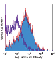
NIH/3T3 cell line stained with biotinylated 55R-170, followed by Sav-PE
| Cat # | Size | Price | Quantity Check Availability | Save | ||
|---|---|---|---|---|---|---|
| 112903 | 50 µg | 72€ | ||||
| 112904 | 500 µg | 216€ | ||||
CD120a is a 55 kD Type I transmembrane protein, also known as Tumor Necrosis Factor Receptor Type I (TNFRI) or p55. It is expressed on a variety of cells at low levels. Resting T cells and erythrocytes express very little to no CD120a. This receptor binds both TNF-α and LT-α (also known as TNF-β). In association with TRADD and RIP, the receptor crosslinking induced by TNF-α or LT-α trimers is critical for signal transduction, leading to apoptosis, NF-kB activation, increased expression of proinflammatory genes, tumor necrosis, and cell differentiation depending on cell type and differentiation state. The 55R-170 antibody has been shown to block in vitro and in vivo receptor signaling initiated by ligand binding.
Product DetailsProduct Details
- Verified Reactivity
- Mouse
- Antibody Type
- Monoclonal
- Host Species
- Armenian Hamster
- Immunogen
- E. coli-expressed extracellular domain of the mouse TNFRI protein
- Formulation
- Phosphate-buffered solution, pH 7.2, containing 0.09% sodium azide.
- Preparation
- The antibody was purified by affinity chromatography, and conjugated with biotin under optimal conditions.
- Concentration
- 0.5 mg/ml
- Storage & Handling
- The antibody solution should be stored undiluted between 2°C and 8°C. Do not freeze.
- Application
-
FC - Quality tested
ELISA Detection - Reported in the literature, not verified in house - Recommended Usage
-
Each lot of this antibody is quality control tested by immunofluorescent staining with flow cytometric analysis. For flow cytometric staining, the suggested use of this reagent is ≤1.0 µg per million cells in 100 µl volume. It is recommended that the reagent be titrated for optimal performance for each application.
- Application Notes
-
Flow Cytometry: For the most successful immunofluorescent staining results, it may be necessary to maximize signal over background by using a relatively bright fluorochrome-antibody conjugate or by using a high sensitivity, three-layer staining technique (e.g., including a biotinylated antibody (Cat. No. 112904) or biotinylated anti-Armenian hamster IgG (Cat. No. 405501) second step, followed by SAv-PE (Cat. No. 405204)).
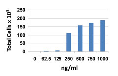
ELISA Detection1: The biotinylated 55R-170 antibody is useful as a detection antibody when paired with the Purified 55R-286 antibody (Cat. No. 113002) as the capture antibody.
Additional reported applications (for the relevant formats) include:immunoprecipitation1, and In vitro and In vivo receptor blocking1. The Ultra-LEAF™Purified antibody (Endotoxin <0.01 EU/µg, Azide-Free, 0.2 µm filtered) is recommended for functional assays (Cat. No. 112905 & 112906). - Application References
-
- Sheehan, K. C., et al. 1995. J. Exp. Med. 181:607.
- RRID
-
AB_313528 (BioLegend Cat. No. 112903)
AB_313529 (BioLegend Cat. No. 112904)
Antigen Details
- Structure
- TNFR superfamily, 55 kD
- Distribution
-
Variety of cell types at low levels
- Function
- Apoptosis, NF-κB activation, inflammation, tumor necrosis, cell differentiation
- Ligand/Receptor
- TNF-α, LT-α (TNF-β)
- Biology Area
- Immunology, Innate Immunity
- Molecular Family
- CD Molecules, Cytokine/Chemokine Receptors
- Antigen References
-
1. Aggarwal, B. B., et al. 1985. Nature 318 665.
2. Chan, F. K.M., et al. 2000. Science 288:2351.
3. Loetscher, H., et al. 1990. Cell 61:351.
4. Rothe, J., et al. 1993. Nature 364:798. - Gene ID
- 21937 View all products for this Gene ID
- UniProt
- View information about CD120a on UniProt.org
Related Pages & Pathways
Pathways


Related FAQs
- How many biotin molecules are per antibody structure?
- We don't routinely measure the number of biotins with our antibody products but the number of biotin molecules range from 3-6 molecules per antibody.
Other Formats
View All CD120a Reagents Request Custom Conjugation| Description | Clone | Applications |
|---|---|---|
| Biotin anti-mouse CD120a (TNF R Type I/p55) | 55R-170 | FC,ELISA Detection |
| Ultra-LEAF™ Purified anti-mouse CD120a (TNF R Type I/p55) | 55R-170 | FC,Block,IP |
Customers Also Purchased
Compare Data Across All Formats
This data display is provided for general comparisons between formats.
Your actual data may vary due to variations in samples, target cells, instruments and their settings, staining conditions, and other factors.
If you need assistance with selecting the best format contact our expert technical support team.
-
Biotin anti-mouse CD120a (TNF R Type I/p55)

NIH/3T3 cell line stained with biotinylated 55R-170, followe... -
Ultra-LEAF™ Purified anti-mouse CD120a (TNF R Type I/p55)

NIH/3T3 cell line stained with purified 55R-170, detected wi...


Follow Us