 Login / Register


- Clone
- MFL3 (See other available formats)
- Regulatory Status
- RUO
- Other Names
- Fas Ligand, FasL, Apo-1 Ligand, CD95 Ligand, TNFSF6
- Isotype
- Armenian Hamster IgG
- Ave. Rating
- Submit a Review
- Product Citations
- publications

-

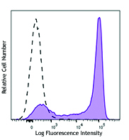
Mouse Fas Ligand transfected cells stained with CD178 (FasL) (clone MFL3) APC (filled histogram) or Armenian hamster IgG APC (open histogram).
| Cat # | Size | Price | Quantity Check Availability | Save | ||
|---|---|---|---|---|---|---|
| 106609 | 25 µg | 96€ | ||||
| 106610 | 100 µg | 214€ | ||||
CD178 is a 40 kD member of the TNF/NGF superfamily also known as Fas ligand, FasL, Apo-1 ligand, and CD95 ligand. Cell surface CD178 is expressed on activated T cells and in testis and eye. CD178 is upregulated in activated T cells upon TCR re-engagement and has been shown to induce autocrine and paracrine T cell death. CD178 expression in the eye and testis has been shown to participate in immune privilege at these sites. CD178 can be cleaved from the surface by metalloproteases, and "soluble" CD178 may block the activities of membrane-bound CD178. CD178 binds to CD95 (Fas) to induce apoptotic cell death implicated in the maintenance of peripheral tolerance. CD178/CD95 interactions have also been implicated in the proliferation of CD8+ cells and neutrophil extravasation, chemotaxis, and survival. The MFL3 antibody recognizes CD178 in a wide array of mouse strains and has been reported to block CD178/CD95 induced apoptosis.
Product DetailsProduct Details
- Verified Reactivity
- Mouse
- Antibody Type
- Monoclonal
- Host Species
- Armenian Hamster
- Immunogen
- B6 mouse FasL cDNA-transfected baby hamster kidney (B6 FasL/BHK) cells
- Formulation
- Phosphate-buffered solution, pH 7.2, containing 0.09% sodium azide.
- Preparation
- The antibody was purified by affinity chromatography and conjugated with APC under optimal conditions.
- Concentration
- 0.2 mg/ml
- Storage & Handling
- The antibody solution should be stored undiluted between 2°C and 8°C, and protected from prolonged exposure to light. Do not freeze.
- Application
-
FC - Quality tested
- Recommended Usage
-
Each lot of this antibody is quality control tested by immunofluorescent staining with flow cytometric analysis. For flow cytometric staining, the suggested use of this reagent is ≤ 0.5 µg per million cells in 100 µl volume. It is recommended that the reagent be titrated for optimal performance for each application.
- Excitation Laser
-
Red Laser (633 nm)
- Application Notes
-
Additional reported applications (for the relevant formats) include: inhibition of the cytotoxicity1-3 and immunofluorescence microscopy4. Fas Ligand is expressed at low density on activated cells. For most successful immunofluorescent staining results, it may be important to maximize signal over background by using a relatively bright fluorochrome-antibody conjugate (Cat. No. 106606) or by using a high sensitivity, three-layer staining technique (e.g., including a biotinylated antibody (Cat. No. 106604) or biotinylated anti-Armenian hamster IgG (Cat. No. 405501) second step, followed by SAv-PE (Cat. No. 405204). The Ultra-LEAF™ purified antibody (Endotoxin < 0.01 EU/µg, Azide-Free, 0.2 µm filtered) is recommended for functional assays (Cat. Nos. 106612).
- Application References
-
- Fuller CL, et al. 1999. J. Immunol. 162:6337.
- Kayagaki N, et al. 1997. P. Natl. Acad. Sci. USA 94:3914.
- Seko Y, et al. 2002. J. Am. Coll.Cardiol. 39:1399.
- Park H, et al. 2005. J. Immunol. 175:7193.
- Lundgvist A, et al. 2009. Blood 113:6120. PubMed
- Oida T, et al. 2011. PLoS ONE 6(4):e18365. (Block) PubMed.
- Oura R, et al. 2013. J. Immunol. 190:578. PubMed.
- Product Citations
-
- RRID
-
AB_2813951 (BioLegend Cat. No. 106609)
AB_2813952 (BioLegend Cat. No. 106610)
Antigen Details
- Structure
- TNF superfamily, 40 kD
- Distribution
-
Activated T cells, spleen, testis, eye
- Function
- Induces apoptosis
- Ligand/Receptor
- Fas (CD95)
- Cell Type
- T cells, Tregs
- Biology Area
- Apoptosis/Tumor Suppressors/Cell Death, Cell Biology, Immunology, Neuroscience
- Molecular Family
- CD Molecules
- Antigen References
-
1. Barclay A, et al. 1997. The Leukocyte Antigen FactsBook Academic Press.
2. Nagata S. 1999. Annu Rev Genet. 33:209.
3. Takahashi T, et al. 1994. Cell. 76:969.
4. Hill LL, et al. 1999. Science. 285:898. - Gene ID
- 14103 View all products for this Gene ID
- UniProt
- View information about CD178 on UniProt.org
Related Pages & Pathways
Pathways


Related FAQs
Other Formats
View All CD178 Reagents Request Custom Conjugation| Description | Clone | Applications |
|---|---|---|
| Biotin anti-mouse CD178 (FasL) | MFL3 | FC |
| PE anti-mouse CD178 (FasL) | MFL3 | FC |
| Purified anti-mouse CD178 (FasL) | MFL3 | FC,ICC |
| APC anti-mouse CD178 (FasL) | MFL3 | FC |
| Ultra-LEAF™ Purified anti-mouse CD178 (FasL) | MFL3 | FC,Block,Apop,ICC |
| TotalSeq™-A1012 anti-mouse CD178 (FasL) | MFL3 | PG |
| TotalSeq™-C1012 anti-mouse CD178 (FasL) | MFL3 | PG |
Customers Also Purchased
Compare Data Across All Formats
This data display is provided for general comparisons between formats.
Your actual data may vary due to variations in samples, target cells, instruments and their settings, staining conditions, and other factors.
If you need assistance with selecting the best format contact our expert technical support team.
-
Biotin anti-mouse CD178 (FasL)

Mouse Fas Ligand transfected cells stained with biotinylated... -
PE anti-mouse CD178 (FasL)

Mouse Fas Ligand transfected cells stained with MFL3 PE -
Purified anti-mouse CD178 (FasL)

Mouse Fas Ligand transfected cells stained with purified MFL... -
APC anti-mouse CD178 (FasL)

Mouse Fas Ligand transfected cells stained with CD178 (FasL)... -
Ultra-LEAF™ Purified anti-mouse CD178 (FasL)
Mouse Fas Ligand transfected cells stained with purified MFL... -
TotalSeq™-A1012 anti-mouse CD178 (FasL)
-
TotalSeq™-C1012 anti-mouse CD178 (FasL)


Follow Us