 Login / Register


- Clone
- Poly28400 (See other available formats)
- Regulatory Status
- RUO
- Other Names
- Glial Fibrillary Acidic Protein
- Previously
-
Covance Catalog# PRB-571C
- Isotype
- Rabbit Polyclonal IgG
- Ave. Rating
- Submit a Review
- Product Citations
- publications

-

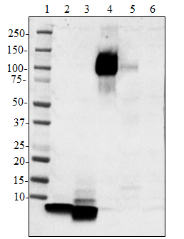
Western blot of anti-GFAP antibody (Poly28400). Lane 1, 4: Molecular weight marker; Lane 2: 20 µg of mouse brain lysate; Lane 3: 20 µg of rat brain lysate; Lane 5: 20 µg of human brain lysate. The blot was incubated with a 1:20000 dilution of the primary antibody overnight at 4°C, followed by incubation with donkey anti-rabbit IgG antibody (Cat. No. 406401). Enhanced chemiluminescence was used as the detection system. -

IHC staining of anti-GFAP antibody (Poly28400) on formalin-fixed paraffin-embedded human brain tissue. Following antigen retrieval using Sodium Citrate H.I.E.R., the tissue was incubated with a 1:1000 dilution of the primary antibody overnight at 4°C, followed by incubation with 2.5 µg/mL of Alexa Fluor® 647 donkey anti-rabbit IgG antibody (Cat. No. 406414) for one hour at room temperature. Nuclei were counterstained with DAPI. The image was captured with a 40X objective. Scale bar: 50 µm -

IHC staining of anti-GFAP antibody (Poly28400) on formalin-fixed paraffin-embedded mouse brain tissue. Following antigen retrieval using Sodium Citrate H.I.E.R., the tissue was incubated with a 1:1000 dilution of the primary antibody overnight at 4°C, followed by incubation with 2.5 µg/mL of Alexa Fluor® 647 donkey anti-rabbit IgG antibody (Cat. No. 406414) for one hour at room temperature. Nuclei were counterstained with DAPI. The image was captured with a 40X objective. Scale bar: 50 µm -

IHC staining of anti-GFAP antibody (Poly28400) on formalin-fixed paraffin-embedded rat brain tissue. Following antigen retrieval using Sodium Citrate H.I.E.R., the tissue was incubated with a 1:1000 dilution of the primary antibody overnight at 4°C, followed by incubation with 2.5 µg/mL of Alexa Fluor® 647 donkey anti-rabbit IgG antibody (Cat. No. 406414) for one hour at room temperature. Nuclei were counterstained with DAPI. The image was captured with a 40X objective. Scale bar: 50 µm
| Cat # | Size | Price | Quantity Check Availability | Save | ||
|---|---|---|---|---|---|---|
| 840001 | 100 µL | 251€ | ||||
Glial Fibrillary Acidic Protein (GFAP) was found to be a member of the 10nm or intermediate filament protein family, specifically the intermediate filament protein family Class III, which also includes peripherin, desmin and vimentin. The GFAP protein runs on gels at ~55kD protein, usually associated with lower molecule weight bands which are thought to be proteolytic fragments and alternate transcripts from the single gene. GFAP is strongly and specifically expressed in astrocytes and certain other astroglia in the central nervous system, in satellite cells in peripheral ganglia, and in non-myelinating Schwann cells in peripheral nerves. In many damage and disease states GFAP expression is heavily upregulated in astrocytes. In addition neural stem cells frequently strongly express GFAP. Antibodies to GFAP are therefore very useful as markers of astrocytic cells and neural stem cells. In addition many types of brain tumor, presumably derived from astrocytic cells, heavily express GFAP. Finally, Alexander's disease was recently shown to be caused by point mutations in protein coding region of the GFAP gene. All forms of Alexander disease are characterized by the presence of Rosenthal fibers, which are GFAP containing cytoplasmic inclusions found in astrocytes.
Product DetailsProduct Details
- Verified Reactivity
- Human, Mouse, Rat
- Antibody Type
- Polyclonal
- Host Species
- Rabbit
- Immunogen
- This antiserum was made with a preparation of recombinant GFAP expressed in bacteria and highly purified. Subsequent boosts were performed with purified GFAP from bovine spinal cord.
- Preparation
- Serum
- Storage & Handling
- Store at -20°C. Upon initial thawing, apportion into working aliquots and store at -20°C. Avoid repeated freeze-thaw cycles to prevent denaturing the antibody. For long-term storage, keep the antibody at -80°C.
- Application
-
WB - Quality tested
IHC-P - Verified - Recommended Usage
-
Each lot of this antibody is quality control tested by western blotting. For western blotting, a dilution range of 1:10000 - 1:20000 is suggested. For immunohistochemistry on formalin-fixed paraffin-embedded tissue sections, a dilution of 1:1000 is suggested. It is recommended that the reagent be titrated for optimal performance for each application.
- Application Notes
-
Multiple protein fragments ranging from 38 to 48 kD have been reported in human CNS lysates resulting from caspase- and calpain-mediated cleavage of GFAP.
- Additional Product Notes
-
This product may contain other non-IgG subtypes.
- Product Citations
-
- RRID
-
AB_2565444 (BioLegend Cat. No. 840001)
Antigen Details
- Structure
- GFAP is a 432 amino acid protein with a molecular mass of ~50 kD.
- Distribution
-
Tissue distribution: GFAP is expressed by numerous cell types of the central nervous system (CNS) including astrocytes, ependymal cells, and Bergmann glia cells (protoplasmic astrocyte). GFAP is expressed in cells lacking fibronectin.
Cellular distribution: Cytoskeleton and cytosol - Function
- GFAP is a class III intermediate filament and a structural constituent of the cytoskeleton. It is a cell-specific marker that is used to distinguish astrocytes from other glial cells during the development of the CNS.
- Cell Type
- Astrocytes
- Biology Area
- Cell Biology, Neuroscience, Neuroscience Cell Markers
- Molecular Family
- Intermediate Filaments
- Antigen References
-
- van Bodegraven EJ, et al. 2019. Glia. 67:1417-1433.
- Pekny M, et al. 2019. Neurosci Lett. 689:45.
- Hol EM, et al. 2017. Cold Spring Harb Perspect Biol. 9(12).
- Gene ID
- 2670 View all products for this Gene ID
- UniProt
- View information about GFAP on UniProt.org
Related Pages & Pathways
Pathways


Related FAQs
Other Formats
View All GFAP Reagents Request Custom Conjugation| Description | Clone | Applications |
|---|---|---|
| Anti-GFAP | Poly28400 | WB,IHC-P |
Customers Also Purchased
Compare Data Across All Formats
This data display is provided for general comparisons between formats.
Your actual data may vary due to variations in samples, target cells, instruments and their settings, staining conditions, and other factors.
If you need assistance with selecting the best format contact our expert technical support team.


Follow Us