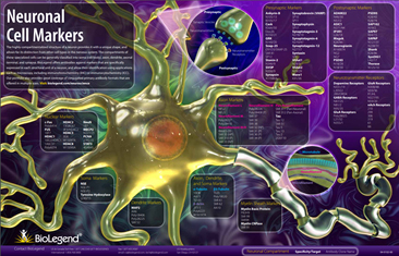
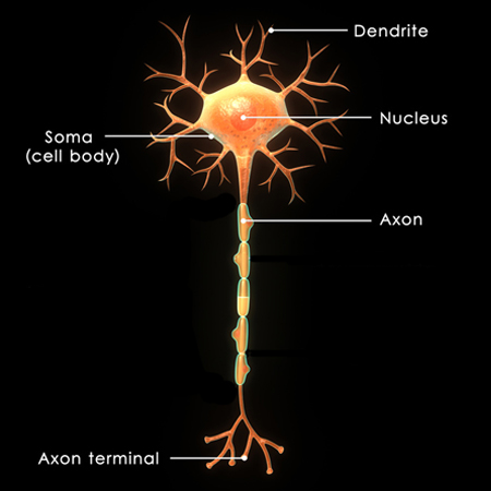
Neurons have highly compartmentalized structures that allow their distinction from other cell types in the nervous system. These unique compartments are distinguishable using specific markers, and are generally classified into soma (cell body), axon, dendrite, and synapse. The use of antibodies for these markers in conjunction with microscopy serves as a powerful method for:

  • Distinguishing neurons from other cell types in the nervous system (e.g. microglia, astrocytes, and oligodendrocytes)
  • Phenotypic and morphological analysis of neurons
  • Assessment of cellular or protein co-localization
  • Visualization of synaptic connections
  • Measurement of protein expression levels
  • Assessment of cell health

Download our complimentary Neuronal Cell Markers Poster.


Immunolabeling of cells (immunocytochemistry or ICC) or tissues (immunohistochemistry or IHC) with antibodies to study neurons is a highly utilized application in Neuroscience mainly due to the availability of a wide range of markers and the relatively low cost for performing and imaging the immunolabeled material. Generally, target detection can be accomplished using chromogenic or fluorescent staining.


Specificity Clone Neuronal Components
    Axon Dendrite Cell Body Nuclear
Enolase 2 NSE-P1      
NeuN 1B7      
NF-H (phospho) SMI 31      
NF-H SMI 32  
NF-H/NF-M (phospho) SMI 35      
MAP2 SMI 52    
Tubulin β 3 TUJ1  

Commonly Used Markers for Neurons


Sections:

β III Tubulin

Enolase 2

Neurofilaments

NeuN

MAP2


BioLegend offers a great number of antibodies validated for IHC and/or ICC. Many of our antibodies are offered in multiple formats and sizes, including small 25 µg vials. Our stringent antibody validation guarantees high product quality and specificity, and the availability of small product sizes allows for hassle-free detection and analysis of neurons.

β III Tubulin


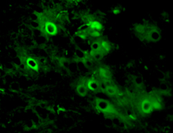
β III Tubulin is a cytoskeletal protein expressed in neurons. Clone TUJ1 is highly reactive to class III β-tubulin, but does not react with β-tubulin found in glial cells. Immunostaining with TUJ1 allows visualization of cell bodies, dendrites, and axons. 

Enolase 2


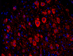
Enolase 2, also known as NSE, is a soluble protein used for identification of neurons and cells of neuronal origin. Staining NSE with clone NSP-P1 can be used to visualize the soma and neuronal processes.

Purified anti-NSE Antibody


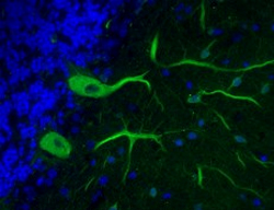
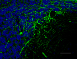
IHC staining of purified anti-NSE antibody (clone NSE-P1, red) on FFPE mouse brain tissue. Nuclei were counterstained with DAPI (blue). 

Neurofilaments


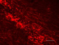
Neurofilaments belong to the intermediate filament family of proteins, and are primarily composed of three subunits; neurofilament light (NF-L), medium (NF-M), and heavy (NF-H). NFs are essential for providing structural support and maintenance of axon caliber. BioLegend has several clones against neurofilaments. Clone SMI 31 detects phosphorylated NF-H and primarily reacts with axons. Clone SMI 32 detects a non-phosphorylated epitope in NF-H and visualizes neuronal cell bodies, axons, and dendrites. Clone SMI 35 reacts with axons and detects phosphorylated forms of NF-M and NF-H.

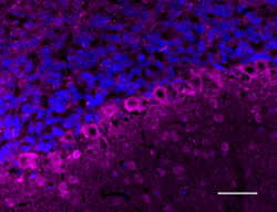
Anti-Neurofilament M (NF-M) Antibody


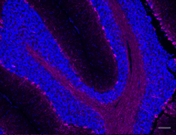
IHC staining of anti-Neurofilament M (NF-M) antibody (Poly28410, magenta) on FFPE rat cerebellum tissue. Nuclei were counterstained with DAPI (blue). 

NeuN


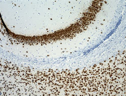
Neuronal Nuclei (NeuN) is also known as Fox3. Staining for NeuN with clone 1B7 reveals strong nuclear staining of a wide range of neuronal cell types. There are some neuronal cells that are not detected by NeuN, such as Purkinje neurons, Golgi cells, and retinal photoreceptor cells.

Purified anti-FOX3 (NeuN) Antibody 


IHC staining of purified anti-FOX3 (NeuN) antibody (clone 1B7) on FFPE mouse brain tissue. The section was counterstained with hematoxylin.

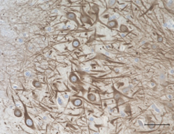
HRP anti-MAP2 Antibody


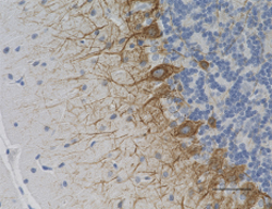
IHC staining of HRP anti-MAP2 antibody (clone SMI 52) on FFPE rat brain tissue. The section was counterstained with hematoxylin and bluing solution. 

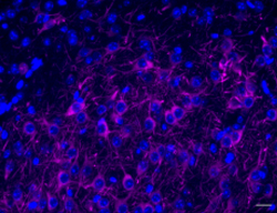
Alexa Fluor® 647 anti-MAP2 Antibody


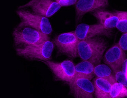
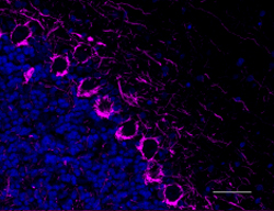
IHC staining of Alexa Fluor® 647 anti-MAP2 antibody (clone SMI 52, magenta) on FFPE mouse brain tissue. Nuclei were counterstained with DAPI (blue). 

ProductsHere

Login / Register
Remember me
Forgot your password? Reset password?
Create an Account