 Login / Register


Tools to Streamline T Cell Research
T cells are an indispensable arm of adaptive immunity and take on distinct immune functions depending on their phenotype1. T cells and their capacity for antigen-directed cytotoxicity have come to play a central role in cancer immunotherapies, treatments that boost the body’s own immune system to target and eliminate malignant tumors. Given the duality of T cells in contributing as cancer medicines and in impeding excessive immunity, T cell biology continues to be a dynamic area of research. Here, we discuss important tools needed for each step of T cell research, from cell isolation, to activation/expansion of T cells, to analysis and characterization of T cell populations.
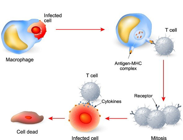
T cells and their immune responses require precise activation involving the integration of three separate signals: its T cell receptor (TCR)-CD3 complex engaging specific peptide antigens on the surface of antigen presenting cells (APCs), the binding of co-stimulatory CD28 receptor to either B7-1 (CD80) or B7-2 (CD86) proteins on APCs, and cytokines that mediate T cell differentiation and expansion2.
To support basic T cell research, or to prime T cell populations for therapeutics and clinical applications, in vitro methods for T cell activation and expansion are now indispensable to generate a large number of functional T cells. First and foremost, you have to start with the right cells. We offer Lymphopure™ to isolate PBMCs and MojoSort™, our magnetic bead-based cell separation system, to isolate T cells and various subtypes. Isolated T cells can then be activated by co-culture with APCs. However, current methods rely on anti-CD3 and anti-CD28 monoclonal antibodies, either soluble, immobilized on plates, or coupled to beads, thus removing the need for feeder cells (APCs) or antigen. Compared to soluble anti-CD3 antibodies, anti-CD3/CD28 beads offer a key advantage in that they are more robust at expanding T cells, with T cell proliferation lasting for a longer time period3.
Our Human CD3/CD28 T Cell Activation Beads act as artificial APCs, designed to mimic in vivo T cell activation by providing consistent and simultaneous signals to the TCR-CD3 complex and the CD28 receptor for complete activation. These superparamagnetic microbeads have a uniform 6 µm diameter, similar size to APCs, and offer robust T cell activation without the need of APC cultures (Figure 1).


Figure 1. (Left) Human CD3+ T cells were isolated from healthy human PBMCs using the MojoSort™ Human CD3 T Cell Isolation Kit (Cat. No. 480021). The CD3+ T cells were expanded with Human CD3/CD28 T Cell Activation Beads. At day 3 post-activation, the beads were removed using a MojoSort™ Magnet (Cat. No. 480019) while the cells remained in culture in the presence of IL-2 (Cat. No. 589102). Our Human CD3/CD28 T Cell Activation Beads stimulate T cells similar to beads from a competitor. (Right) PD-1 expression at day 5 of culture of human CD3+ T cells stimulated with Human CD3/CD28 T Cell Activation Beads. At day of harvest, cells were stained with anti-human CD3 (clone SK7) APC (allophycocyanin) and anti-human CD279 (PD-1) (clone NAT105) Brilliant Violet 421™ and were analyzed by flow cytometry. Cell debris, dead cells, and doublets were excluded from analysis.
For our partners who are ready to translate research to medicines and therapeutics, know that BioLegend is a leader in the field accelerating research and discovery by providing high-quality GMP recombinant proteins, functional antibodies, and cell culture reagents for use throughout the bioprocessing workflow. Learn about our monoclonal antibodies GMP Ultra-LEAF™ Purified anti-human CD3 SF Antibody (clone OKT3) and GMP Ultra-LEAF™ Purified anti-human CD28 SF Antibody (clone S20013F), produced under serum-free (SF) conditions from start to finish. We source the highest quality raw materials to manufacture these SF antibodies, thus removing the risk of using serum while providing additional control, consistency, and safety for T cell-based workflows.
After T cell activation, signaling from key cytokines and growth factors (such IL-2, IL-4, IL-6, TGF-β, TNF-α, and IFN-γ) help drive T cells to differentiate. Research on the nature and function of these T cell subtypes is greatly facilitated by examining expression of cell surface markers and cytokine secretion profiles (Figure 2). Explore our interactive page on T helper cell phenotypes that helps you locate cell surface markers, differentiation cytokines, and effector cytokines for the T cell subtype of interest. We offer an extensive portfolio of recombinant cytokines and growth factors, each of which undergoes rigorous quality testing and verification of bioactivity using functional assays.
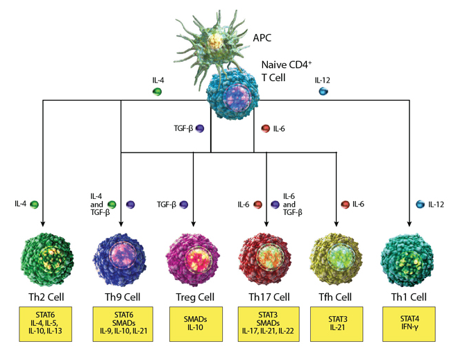
Figure 2. Pathways of T helper cell differentiation in response to interleukin signaling.
For cell therapeutics, biomanufacturers can rely on our GMP recombinant proteins, ideal for research or ex vivo cell bioprocessing. These proteins are manufactured and tested in a dedicated GMP facility that is ISO 13485:2016 and MDSAP certified, monitored through independent QA oversight.
For more ways to phenotype T cell subtypes, utilize our Essential Markers resource for a list of phenotypic markers commonly documented in literature to identify the cell type of interest. After identifying the markers you want, start building the multicolor flow panel you need to study the complexity – both functionally and phenotypically – of T cell populations. We also offer detailed flow cytometry protocols, such as use of intracellular flow cytometry to detect effector molecules (including cytokines and chemokines) and signaling molecules (such as transcription factors and phosphorylated proteins). To get a complete profile of T cells, it is useful to measure their secreted cytokines. Use ELISA and LEGEND MAX™ to quantitate levels of single cytokines secreted by T cells or LEGENDplex™ for multiplexed flow-based immunoassays that measure multiple cytokines in the same sample. We offer pre-built panels for T helper cells, CD8/NK, and other cell types.
We understand the central role T cells play in cell-mediated immunity and how important the study of T cells is to help decipher complex pathologies such as cancer and autoimmunity. At BioLegend, our scientists develop tools for each step of T cell research. By establishing the unique phenotypic signature carried by specific immune cells, researchers can study how immune cells respond to different environments, such as disease state, and help uncover key mechanisms that underly diseases such as cancer. Want to learn more? Read our blog on the rich history of T cell research and Nature’s Milestones celebration of pivotal T cell studies and their potential to advance human health.
References
- Zhu, Jinfang et al. “Differentiation of effector CD4 T cell populations (*).” Annual review of immunology vol. 28 (2010): 445-89. doi:10.1146/annurev-immunol-030409-101212. PubMed.
- Xia, Fan et al. “TCR and CD28 Concomitant Stimulation Elicits a Distinctive Calcium Response in Naive T Cells.” Frontiers in immunology vol. 9 2864. 4 Dec. 2018, doi:10.3389/fimmu.2018.02864. PubMed.
- Li, Yixin, and Roger J Kurlander. “Comparison of anti-CD3 and anti-CD28-coated beads with soluble anti-CD3 for expanding human T cells: differing impact on CD8 T cell phenotype and responsiveness to restimulation.” Journal of translational medicine vol. 8 104. 26 Oct. 2010, doi:10.1186/1479-5876-8-104. PubMed.


Follow Us