 Login / Register


- Clone
- Poly19010 (See other available formats)
- Regulatory Status
- RUO
- Other Names
- Paired box protein Pax-2, paired box homeotic gene 2, Opdc, optic disc coloboma
- Previously
-
Covance Catalog# PRB-276P
- Isotype
- Rabbit Polyclonal IgG
- Ave. Rating
- Submit a Review
- Product Citations
- publications

-

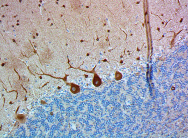
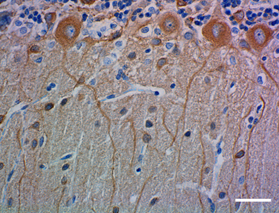
Pax2 staining on normal human kidney tissue, demonstrating strong nuclear staining of epithelial cells in some renal tubules. -

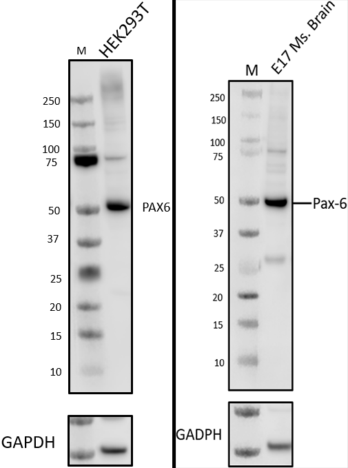
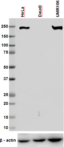
Western blot analysis of cell lysates from MCF-7 (low expression negative control) and Daudi (positive control) cells using Pax-2 rabbit primary antibody (Clone Poly19010,1:1000 dilution, 1 µg/ml) and HRP Donkey anti-rabbit secondary antibody (Cat. No. 406401, 1:3000 dilution). Direct-Blot™ HRP anti-GAPDH (Cat. No. 649203) was used as a loading control (1:1000 dilution).
| Cat # | Size | Price | Quantity Check Availability | Save | ||
|---|---|---|---|---|---|---|
| 901002 | 25 µL | £61 | ||||
| 901001 | 200 µL | £254 | ||||
Pax genes are expressed during embryogenesis in a temporally and spatially restricted manner in the developing neural tube. Pax-2 has been identified to be involved in kidney and optic nerve development. It belongs to the Pax group 2, including Pax-5 and Pax-8. Paired box (Pax) proteins are tissue specific transcription factors that contain a paired domain and typically a partial or complete homeodomain.
Product DetailsProduct Details
- Verified Reactivity
- Human
- Reported Reactivity
- Mouse, Chicken, Frog, Zebrafish
- Antibody Type
- Polyclonal
- Host Species
- Rabbit
- Immunogen
- This Pax-2 antibody was generated against a recombinant protein containing 22kD of the Pax-2 sequence corresponding to amino acids 188-385.
- Formulation
- Phosphate-buffered solution + 0.03% Thimerosal.
- Preparation
- The antibody was purified by affinity chromatography.
- Concentration
- 1.0 mg/ml
- Storage & Handling
- The antibody solution should be stored undiluted between 2°C and 8°C. Please note the storage condition for this antibody has been changed from -20°C to between 2°C and 8°C. You can also check your vial or your CoA to find the most accurate storage condition for this antibody.
- Application
-
IHC-P - Quality tested
WB - Verified
IP - Reported in the literature, not verified in house - Recommended Usage
-
Each lot of this antibody is quality control tested by immunohistochemical staining.
The optimal working dilution should be determined for each specific assay condition.
• WB: 1-2 µg/ml (1:500-1:1000)
• Gel shifts: 1:10
• IHC: 1:100
Tissue Sections: Formalin-fixed, paraffin-embedded tissues, acetone or PFA fixed frozen sections
Pretreatment: For optimal staining, the sections should be pretreated with an antigen unmasking solution such as Retrieva-All 1 (Cat. No. 927901).
Incubation: 20 minutes at room temperature
Staining Localization: nuclear - Application Notes
-
This antibody is effective in immunoblotting (WB), immunohistochemistry (IHC), immunopurification and DNA band supershift experiments.
*Predicted MW = 44 kD
Positive Control Tissue: normal kidney or renal adenocarcinoma
This antibody recognizes Pax-2A and Pax-2B (two products of the Pax-2 gene). When linked to agarose beads, it has been used to immunoprecipitate DNA sequences recognized by Pax-2.
This product may contain other non-IgG subtypes. -
Application References
(PubMed link indicates BioLegend citation) -
- Hammes A, et al. 2001. Cell. 106:319.
- Phelps DE, Dressler GR. 1996. J Biol Chem. 271:7978.
- Rothenpieler UW, Dressler GR. 1993. Development. 119:711
- Dressler GR, et al. 1993. Nature. 362:65
- Dressler GR, Douglass EC. 1992. Proc Natl Acad Sci USA. 89:1179
- Puschel AW, et al. 1992. Mech Dev. 38:197
- Riley BB, et al. 1999. Development. 126:5669. (IHC)
- Product Citations
-
- RRID
-
AB_2734656 (BioLegend Cat. No. 901002)
AB_2565001 (BioLegend Cat. No. 901001)
Antigen Details
- Biology Area
- Cell Biology, Neuroscience, Signal Transduction, Synaptic Biology, Transcription Factors
- Molecular Family
- Nuclear Markers
- Gene ID
- 5076 View all products for this Gene ID
- UniProt
- View information about Pax-2 on UniProt.org

Related FAQs
Other Formats
View All Pax-2 Reagents Request Custom Conjugation| Description | Clone | Applications |
|---|---|---|
| Purified anti-Pax-2 | Poly19010 | IHC-P,WB,IP |
Customers Also Purchased
Compare Data Across All Formats
This data display is provided for general comparisons between formats.
Your actual data may vary due to variations in samples, target cells, instruments and their settings, staining conditions, and other factors.
If you need assistance with selecting the best format contact our expert technical support team.


Follow Us