 Login / Register


- Clone
- 4D1 (See other available formats)
- Regulatory Status
- RUO
- Other Names
- NF-κB p50, nuclear factor kappa light chain enhancer in B cells p50, NF-κB1
- Isotype
- Mouse IgG1, κ
- Ave. Rating
- Submit a Review
- Product Citations
- publications

-

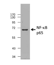
Total cell lysates (15 µg protein) from Jurkat (Lane 1) cells were resolved by electrophoresis (4-20% Tris-Glycine gel), transferred to nitrocellulose, and probed with 0.025 µg/mL (1:20,000 dilution) Purified anti-NF-κB p50 Antibody, clone 4D1 (upper panel). Proteins were visualized by chemiluminescence detection using a 1:3000 diluted goat anti-mouse-IgG secondary antibody conjugated with HRP for anti-NF-κB p50 Antibody , or 1:5000 diluted Direct-Blot HRP anti-β-Actin Antibody, clone 2F1-1 (lower panel) as a loading control. Lane M: Molecular weight ladder. -

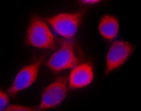
A549 cells were unstimulated (left panel) or treated with 20 ng/mL TNF-α (Cat. No. 717904, right panel) for 16 hours. Cells were then fixed with 4% paraformaldehyde for 10 minutes, permeabilized with 0.5% Triton X-100 for 10 minutes, and blocked with 5% FBS for 60 minutes. Cells were then intracellularly stained with 2.0 µg/mL (1:250 dilution) of Purified anti-NF-κB Antibody, clone 4D1, overnight at 4°C, after which proteins were visualized with Alexa Fluor® 594 Goat anti-mouse IgG Antibody (Cat. No. 405326) at 2.0 µg/mL. Nuclei were counterstained with DAPI, and the image was captured with a 60X objective.
| Cat # | Size | Price | Quantity Check Availability | Save | ||
|---|---|---|---|---|---|---|
| 616701 | 25 µg | £70 | ||||
| 616702 | 100 µg | £150 | ||||
NF-κB p50 (nuclear factor kappa light chain enhancer in B cells p50, NF-κB1) is a member of the Rel/dorsal family. NF-κB is a transcription factor, originally identified as an activator of kappa light chain in B cells. NF-κB p50, together with another NF-κB subunit p65, binds to IκB inhibitor in cytoplasm as an inactive form. Phosphorylation of IκB releases NF-κB p50 and p65. Active form of NF-κB p50 and p65 then translocate to the nucleus and regulate target gene expression. Bacterial products and stimulation of a variety of receptors lead to NF-κB signal pathway activation. This 4D1 monoclonal antibody recognizes p105 (precursor of p50) and p50 (active form). 4D1 has been validated for Western blotting.
Product DetailsProduct Details
- Verified Reactivity
- Human
- Antibody Type
- Monoclonal
- Host Species
- Mouse
- Immunogen
- recombinant full-length human NF-κB p50
- Formulation
- This antibody is provided in phosphate-buffered solution, pH 7.2, containing 0.09% sodium azide. Final antibody concentration is 0.5 mg/ml.
- Preparation
- The antibody was purified by affinity chromatography.
- Concentration
- 0.5 mg/ml
- Storage & Handling
- Upon receipt, store between 2°C and 8°C.
- Application
-
WB - Quality tested
ICC - Reported in the literature, not verified in house - Recommended Usage
-
Each lot of this antibody is quality control tested by Western blotting. For Western blotting, the suggested use of this reagent is 0.025 - 0.5 µg per ml. For immunocytochemistry, a concentration range of 2.0 μg/mL is recommended. It is recommended that the reagent be titrated for optimal performance for each application.
- Application Notes
-
Additional reported applications (for the relevant formats) include: immunofluorescence1 on the Amnis ImageStream 100 Imaging Flow Cytometer.
This clone is not recommended for ChIP (Chromatin Immunoprecipitation) assays (as determined by in-house testing). -
Application References
(PubMed link indicates BioLegend citation) - Product Citations
-
- RRID
-
AB_315855 (BioLegend Cat. No. 616701)
AB_315856 (BioLegend Cat. No. 616702)
Antigen Details
- Structure
- Member of the Rel/dorsal family. NF-κB p50 is an active form, derived from NF-κB p105, a precursor form.
- Distribution
-
Ubiquitously expressed
- Function
- A transcription factor, originally identified as an activator of kappa light chain in B cells
- Interaction
- Interacts with NF-κB p65 and IκB
- Biology Area
- Cell Biology, Immunology, Transcription Factors
- Molecular Family
- Nuclear Markers
- Antigen References
-
1. Baldwin AS, et al. 1996. Annu. Rev. Immunol. 14:649 (review).
2. Chen F, et al. 1999. Clin. Chem. 45:7 (review). - Regulation
- NF-κB p50, together with another NF-κB subunit p65, binds to IκB inhibitor in cytoplasm as an inactive form. Phosphorylation of IκB releases NF-κB p50 and p65. Active form of NF-κB p50 and p65 then translocate to the nucleus and regulate target gene expression. Bacterial products and stimulation of a variety of receptors lead to NF-κB signal pathway activation.
- Gene ID
- 4790 View all products for this Gene ID
- UniProt
- View information about NF-kappaB p50 on UniProt.org

Related FAQs
Other Formats
View All NF-κB p50 Reagents Request Custom Conjugation| Description | Clone | Applications |
|---|---|---|
| Purified anti-NF-κB p50 | 4D1 | WB,ICC |
Customers Also Purchased
Compare Data Across All Formats
This data display is provided for general comparisons between formats.
Your actual data may vary due to variations in samples, target cells, instruments and their settings, staining conditions, and other factors.
If you need assistance with selecting the best format contact our expert technical support team.


Follow Us