 Login / Register


- Clone
- M3/38 (See other available formats)
- Regulatory Status
- RUO
- Other Names
- Galectin-3, Mac-2, Gal-3, RL-29, galactose-specific lectin 3, CBP-35, L-34, εBP
- Isotype
- Rat IgG2a, κ
- Ave. Rating
- Submit a Review
- Product Citations
- publications

-

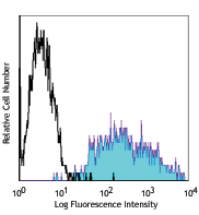
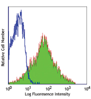
BALB/c peritoneal macrophages stained with M3/38 purified, followed by anti-rat IgG FITC -

HeLa cells were stained with purified anti-Galectin 3 (M3/38) antibody, followed by staining with DyLight™ 594 conjugated goat anti-mouse IgG (red) antibody. Actin filaments were labeled in green. Nuclei were stained with DAPI (blue). -

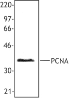
Immunoprecipitation/Western Blot analysis using anti‐mouse/human Mac‐2 (Galectin‐3) Antibody (clone M3/38). 150 µg of HeLa cell lysates were tested at a protein concentration of 1 µg/µL for each sample. Lane 1 was immunoprecipitated with a control antibody and lane 2 was immunoprecipitated with anti‐mouse/human Mac‐2 (Galectin‐3) Antibody (clone M3/38). Western blot analysis was performed using anti‐mouse/human Mac‐2 antibody (clone Gal397). -

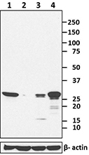
Total cell lysate from MCF7 cells (lane 1, 15 µg), Jurkat cells (lane 2, 15 µg), 3T3-L1 (lane 3, 15 µg) and Raw264.7 (lane 4, 15 µg) were resolved by electrophoresis (4-20% Tris-Glycine gel), transferred to nitrocellulose, and probed with purified anti-mouse/human Mac-2 (Galectin-3) antibody (clone M3/38). Proteins were visualized using an HRP Goat anti-rat IgG Antibody and chemiluminescence detection. Direct-Blot™ HRP anti-β-actin antibody (clone 2F1-1) was used as a loading control.
| Cat # | Size | Price | Quantity Check Availability | Save | ||
|---|---|---|---|---|---|---|
| 125401 | 50 µg | £50 | ||||
| 125402 | 500 µg | £177 | ||||
Galectins, a family of carbohydrate binding proteins (lectins) have been implicated in inflammation and cancer. All galectins bind lactose and other beta-galactosides but differ in their affinity for more complex saccharides.
Product DetailsProduct Details
- Verified Reactivity
- Mouse, Human
- Antibody Type
- Monoclonal
- Host Species
- Rat
- Immunogen
- Raised against galectin-3 of mouse origin
- Formulation
- Phosphate-buffered solution, pH 7.2, containing 0.09% sodium azide.
- Preparation
- The antibody was purified by affinity chromatography.
- Concentration
- 0.5 mg/mL
- Storage & Handling
- The antibody solution should be stored undiluted between 2°C and 8°C.
- Application
-
FC - Quality tested
ICC, WB, IP - Verified
IHC, ELISA - Reported in the literature, not verified in house - Recommended Usage
-
Each lot of this antibody is quality control tested by immunofluorescent staining with flow cytometric analysis. For flow cytometric staining, the suggested use of this reagent is ≤ 0.25 µg per 106 cells in 100 µL volume or 100 µL of whole blood. It is recommended that the reagent be titrated for optimal performance for each application.
- Application Notes
-
Additional reported applications (for the relevant formats) include: immunohistochemical staining of paraffin-embedded tissue sections3-6, Western blotting2, immunoprecipitation1,2, immunofluorescence7,8, and ELISA9.
Clone M3/38 has been reported to recognize residues 48-100 in the amino-terminal domain of galectin-37.
-
Application References
(PubMed link indicates BioLegend citation) -
- Ho MK. and Springer TA. 1982. J. Immunol. 128:1221. (FC, IP)
- Rosenberg I, et al. 1991. J. Biol. Chem. 266:18731. (WB, IP)
- Evans CE, et al. 2010. Arterioscler Vasc Biol. (IHC) PubMed
- Jacob N, et al. 2011. J. Immunol. 186:4984. (IHC) PubMed
- Li X, et al. 2011. Am J Physiol Heart Circ Physiol. 301:1932. (IHC) PubMed
- Chao C, et al. 2012. Clin Cancer Res. 18:4702. (IHC) PubMed
- Melo FH, et al. 2011. PLoS One. 6:e29313. (IF)
- Usategui A, et al. 2013. Ann Rheum Dis. 72:2018. PubMed (IF)
- Mey A, et al. 1996. J. Immunol. 156:1572. (ELISA)
- Reales E, et al. 2015. J Cell Sci. 128:2261. PubMed
- Product Citations
-
- RRID
-
AB_1134237 (BioLegend Cat. No. 125401)
AB_1134238 (BioLegend Cat. No. 125402)
Antigen Details
- Biology Area
- Cell Biology, Immunology, Innate Immunity
- Molecular Family
- Adhesion Molecules
- Antigen References
-
1. Ho MK. and Springer TA. 1982. J. Immunol. 128:1221.
2. Rosenberg I, et al. 1991. J. Biol. Chem. 266:18731. - Gene ID
- 16854 View all products for this Gene ID 3958 View all products for this Gene ID
- UniProt
- View information about Mac-2 on UniProt.org

Related FAQs
Other Formats
View All Mac-2 Reagents Request Custom Conjugation| Description | Clone | Applications |
|---|---|---|
| Purified anti-mouse/human Mac-2 (Galectin-3) | M3/38 | FC,ICC,WB,IP,IHC,ELISA |
| Biotin anti-mouse/human Mac-2 (Galectin-3) | M3/38 | FC,IHC |
| PE anti-mouse/human Mac-2 (Galectin-3) | M3/38 | FC |
| Alexa Fluor® 647 anti-mouse/human Mac-2 (Galectin-3) | M3/38 | FC,IHC-P |
| Alexa Fluor® 488 anti-mouse/human Mac-2 (Galectin-3) | M3/38 | FC |
| Alexa Fluor® 594 anti-mouse/human Mac-2 (Galectin-3) | M3/38 | IHC-F |
| Brilliant Violet 421™ anti-mouse/human Mac-2 (Galectin-3) | M3/38 | IHC-F |
| PE/Cyanine7 anti-mouse/human Mac-2 (Galectin-3) | M3/38 | FC |
| APC anti-mouse/human Mac-2 (Galectin-3) | M3/38 | FC |
| TotalSeq™-A0895 anti-mouse/human Mac-2 (Galectin-3) | M3/38 | PG |
| TotalSeq™-C0895 anti-mouse/human Mac-2 (Galectin-3) | M3/38 | PG |
| TotalSeq™-B0895 anti-mouse/human Mac-2 (Galectin-3) | M3/38 | PG |
Customers Also Purchased
Compare Data Across All Formats
This data display is provided for general comparisons between formats.
Your actual data may vary due to variations in samples, target cells, instruments and their settings, staining conditions, and other factors.
If you need assistance with selecting the best format contact our expert technical support team.
-
Purified anti-mouse/human Mac-2 (Galectin-3)

BALB/c peritoneal macrophages stained with M3/38 purified, f... 
HeLa cells were stained with purified anti-Galectin 3 (M3/38... 
Total cell lysate from MCF7 cells (lane 1, 15 µg), Jurkat ce... 
Immunoprecipitation/Western Blot analysis using anti‐mouse/h... -
Biotin anti-mouse/human Mac-2 (Galectin-3)

BALB/c peritoneal marcophages stained with M3/38 biotin, fol... -
PE anti-mouse/human Mac-2 (Galectin-3)

BALB/c peritoneal marcophages stained with M3/38 PE -
Alexa Fluor® 647 anti-mouse/human Mac-2 (Galectin-3)

Thioglycolate-elicited BALB/c mouse peritoneal macrophages s... 
Human paraffin-embedded intestine tissue slices were prepare... -
Alexa Fluor® 488 anti-mouse/human Mac-2 (Galectin-3)

Thioglycolate-elicited BALB/c mouse peritoneal macrophages w... 
-
Alexa Fluor® 594 anti-mouse/human Mac-2 (Galectin-3)

C57BL/6 mouse frozen colon section was fixed with 4% parafor... -
Brilliant Violet 421™ anti-mouse/human Mac-2 (Galectin-3)

C57BL/6 mouse frozen colon section was fixed with 4% parafor... -
PE/Cyanine7 anti-mouse/human Mac-2 (Galectin-3)

Thioglycolate-elicited BALB/c mouse peritoneal macrophages w... -
APC anti-mouse/human Mac-2 (Galectin-3)

Thioglycolate-elicited BALB/c mouse peritoneal macrophages w... -
TotalSeq™-A0895 anti-mouse/human Mac-2 (Galectin-3)
-
TotalSeq™-C0895 anti-mouse/human Mac-2 (Galectin-3)
-
TotalSeq™-B0895 anti-mouse/human Mac-2 (Galectin-3)


Follow Us