 Login / Register


- Clone
- APA1/1 (See other available formats)
- Regulatory Status
- RUO
- Other Names
- CD3e molecule, epsilon (CD3-TCR complex), CD3e antigen, CD3 epsilon
- Isotype
- Mouse IgG1, κ
- Ave. Rating
- Submit a Review
- Product Citations
- publications

-

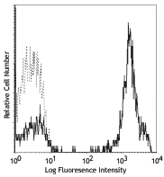
Human peripheral blood lymphocytes were fixed and permeabilized with 70% ethanol, followed by intracellular staining with purified CD3e (clone APA1/1, filled histogram) or mouse IgG1, κ isotype control (open histogram) followed by anti-mouse IgG PE. -

Western blot analysis of extracts from Jurkat (lane 1) and PBMC untreated (lane 2) and PBMC activated with CD3/CD28(lane 3) cells using anti-CD3ε (activation epitope) antibody (clone APA1/1). β-actin (poly6221) was used as loading control. -

Human paraffin-embedded tonsil tissue slices were prepared with a standard protocol of deparaffination and rehydration. Antigen retrieval was done with Tris buffer 1X pH 8.5 at 95°C for 40 minutes. Tissue was washed with PBS/ 0.05% Tween20 twice for five minutes, blocked with 5% FBS and 0.2% gelatin for 30 minutes. Then, the tissue was stained with 10 µg/mL of purified anti-human CD3ε (clone APA1/1) at 4°C overnight. On the next day, the tissue was washed twice with PBS and stained with goat anti-mouse secondary antibody (clone Poly4053) Alexa Fluor® 594 (red) for two hours at room temperature. The nuclei were counter staining with DAPI (blue). The image was captured with a 10X objective. -

Human paraffin-embedded spleen tissue slices were prepared with a standard protocol of deparaffination and rehydration. Antigen retrieval was done with Tris buffer 1X pH 8.5 at 95°C for 40 minutes. Tissue was washed with PBS/ 0.05% Tween20 twice for five minutes, blocked with 5% FBS and 0.2% gelatin for 30 minutes. Then, the tissue was stained with 10 µg/mL of purified anti-human CD3ε (clone APA1/1) and anti-human IgD (clone W18340A) at 4°C overnight. On the next day, the tissue was washed twice with PBS and stained with goat anti-mouse secondary antibody (clone Poly4053) Alexa Fluor® 594 (red) and anti-rat secondary antibody (clone Poly4054) Alexa Fluor® 647 (green) for two hours at room temperature. The nuclei were counter staining with DAPI (blue). The image was captured with a 10X objective.
| Cat # | Size | Price | Quantity Check Availability | Save | ||
|---|---|---|---|---|---|---|
| 362701 | 50 µg | £45 | ||||
| 362702 | 500 µg | £177 | ||||
The CD3ε chain of the CD3-TCR complex is a type I membrane protein that undergoes conformational change following ligation of the TCR-CD3 complex. This conformational change leads to exposure of the proline rich sequence in CD3ε and results in recruitment of Nck. It is expressed by all mature T cells and forms a heteromeric complex with CD3 α/β or γ/δ and ζ chains. It is important in T cell development, and deficiency of this gene has been associated with severe immunodeficiency.
Product DetailsProduct Details
- Verified Reactivity
- Human, Mouse
- Antibody Type
- Monoclonal
- Host Species
- Mouse
- Immunogen
- Purified denatured CD3 proteins from human thymus
- Formulation
- Phosphate-buffered solution, pH 7.2, containing 0.09% sodium azide.
- Preparation
- The antibody was purified by affinity chromatography.
- Concentration
- 0.5 mg/mL
- Storage & Handling
- The antibody solution should be stored undiluted between 2°C and 8°C.
- Application
-
ICFC - Quality tested
WB, IHC-P - Verified
in vivo cell tracking - Reported in the literature, not verified in house
ICC, IHC, IP - Reported by the developer, not verified in house - Recommended Usage
-
Each lot of this antibody is quality control tested by immunofluorescent staining with flow cytometric analysis. For flow cytometric staining, the suggested use of this reagent is ≤ 0.5 µg per million cells in 100 µL volume. For Western blotting, the suggested use of this reagent is 1.0 - 4.0 µg per mL. For immunohistochemistry, a concentration range of 5.0 - 10.0 µg/mL is suggested. It is recommended that the reagent be titrated for optimal performance for each application.
- Application Notes
-
Clone APA1/1 recognizes an activation-dependent intracellular epitope of the CD3ε chain. Additional reported applications (for the relevant formats of this clone) include: immunofluorescent staining of paraformaldehyde fixed sections1, in vivo tracking of T cells 2, and Western blotting3. The LEAF purified format is recommended for in vivo studies.
-
Application References
(PubMed link indicates BioLegend citation) -
- Salmerón A, et al. 1991. J. Immunol. 147:3047. (ICFC, IF)
- Risueño RM, et al. 2005. Blood. 106:601. (IF, in vivo tracking, Mouse Reactivity)
- Risueño RM, et al. 2008. PLos One. 3:e1747. (WB)
- Gil D, et al. 2005. J. Exp. Med. 201:517.
- Risueño RM, et al. 2006. Proc. Natl. Acad. Sci. USA. 103:9625.
- Product Citations
-
- RRID
-
AB_2563713 (BioLegend Cat. No. 362701)
AB_2563714 (BioLegend Cat. No. 362702)
Antigen Details
- Structure
- Type I membrane protein, part of the TCR-CD3 complex. 207 amino acids, predicted molecular weight of 23 kD.
- Distribution
-
Mature T cells.
- Function
- Important role in T cell development and in linking antigen recognition with intracellular signaling pathways.
- Interaction
- Interacts with Nck.
- Ligand/Receptor
- MHC antigen.
- Cell Type
- T cells
- Biology Area
- Cell Adhesion, Cell Biology, Immunology, Signal Transduction
- Molecular Family
- CD Molecules, TCRs
- Antigen References
-
1. Savage PD, et al. 1988. Cytogenet. Cell. Genet. 49:289.
2. DeJarnette JB, et al. 1998. Proc. Natl. Acad. Sci USA. 95:14909.
3. Wang B, et al. 1999. J. Immunol. 162:88.
4. Szymczak AL, et al. 2005. J. Immunol. 175:270.
5. Borroto A, et al. 2014. J. Immunol. 192:2042. - Gene ID
- 916 View all products for this Gene ID
- UniProt
- View information about CD3epsilon on UniProt.org


Related FAQs
Other Formats
View All CD3ε Reagents Request Custom Conjugation| Description | Clone | Applications |
|---|---|---|
| Purified anti-human/mouse CD3ε (activation epitope) | APA1/1 | ICFC,WB,IHC-P,in-vivo Cell Tracking,ICC,IHC,IP |
| PE anti-human/mouse CD3ε (activation epitope) | APA1/1 | ICFC |
| APC anti-human/mouse CD3ε (activation epitope) | APA1/1 | ICFC |
Customers Also Purchased
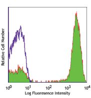
Compare Data Across All Formats
This data display is provided for general comparisons between formats.
Your actual data may vary due to variations in samples, target cells, instruments and their settings, staining conditions, and other factors.
If you need assistance with selecting the best format contact our expert technical support team.
-
Purified anti-human/mouse CD3ε (activation epitope)

Human peripheral blood lymphocytes were fixed and permeabili... 
Western blot analysis of extracts from Jurkat (lane 1) and P... 
Human paraffin-embedded tonsil tissue slices were prepared w... 
Human paraffin-embedded spleen tissue slices were prepared w... -
PE anti-human/mouse CD3ε (activation epitope)

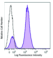
Human peripheral blood lymphocytes were fixed and permeabili... -
APC anti-human/mouse CD3ε (activation epitope)

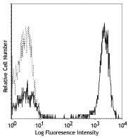
Human peripheral blood lymphocytes were fixed and permeabili...


Follow Us