 Login / Register


- Clone
- WM59 (See other available formats)
- Regulatory Status
- RUO
- Workshop
- V P025
- Other Names
- PECAM-1, EndoCAM
- Isotype
- Mouse IgG1, κ
- Ave. Rating
- Submit a Review
- Product Citations
- publications

-

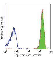
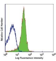
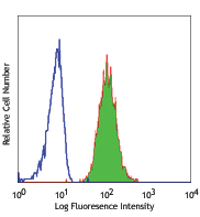
Human peripheral blood lymphocytes, monocytes and granulocytes stained with purified WM59, then detected with anti-mouse IgG FITC -

-

HUVEC cells were fixed with 1% paraformaldehyde (PFA) and blocked with 5% fetal bovine serum for 30 minutes at room temperature. Then the cells were stained with 10 µg/ml of purified anti-CD31 (clone WM59) overnight, followed by 2.5 µg/ml DyLight™ 594 anti-mouse IgG for 2 hours at 4°C (red). Nuclei were counterstained with DAPI (blue). The image was captured by 20X objective.
| Cat # | Size | Price | Quantity Check Availability | Save | ||
|---|---|---|---|---|---|---|
| 303101 | 25 µg | £37 | ||||
| 303102 | 100 µg | £70 | ||||
CD31 is a 130-140 kD type I transmembrane glycoprotein also known as platelet endothelial cell adhesion molecule-1 (PECAM-1) or Endocam. It is expressed on monocytes, platelets, granulocytes, endothelial cells and lymphocyte subsets. CD31 has been reported to bind CD38 and be involved in wound healing, angiogenesis, and cellular migration in an inflammatory situation.
Product DetailsProduct Details
- Verified Reactivity
- Human, Cynomolgus, Rhesus
- Reported Reactivity
- African Green, Baboon
- Antibody Type
- Monoclonal
- Host Species
- Mouse
- Formulation
- Phosphate-buffered solution, pH 7.2, containing 0.09% sodium azide.
- Preparation
- The antibody was purified by affinity chromatography.
- Concentration
- 0.5 mg/ml
- Storage & Handling
- The antibody solution should be stored undiluted between 2°C and 8°C.
- Application
-
FC - Quality tested
ELISA Capture, ICC - Verified
IHC - Reported in the literature, not verified in house - Recommended Usage
-
Each lot of this antibody is quality control tested by immunofluorescent staining with flow cytometric analysis. For flow cytometric staining, the suggested use of this reagent is ≤ 1.0 µg per million cells in 100 µl volume. For ELISA Capture applications, a concentration of 1.0 µg/mL is recommended. To obtain a linear standard curve, serial dilutions of CD31 recombinant protein ranging from 10 to 0.156 ng/mL is recommended for each ELISA plate. For immunocytochemistry, a concentration range of 2.5 - 10 μg/ml is recommended. It is recommended that the reagent be titrated for optimal performance for each application.
- Application Notes
-
Clone WM59 has been reported to recognize the D2 extracellular portion of CD31.
Additional reported applications (for the relevant formats) include: immunofluorescence microscopy2, immunohistochemical staining of acetone-fixed frozen tissue sections8, blocking of platelet aggregation3, and spatial biology (IBEX)11,12. Clone WM59 is not recommended for immunohistochemical staining of formalin-fixed paraffin-embedded sections. The Ultra-LEAF™ purified antibody (Endotoxin < 0.01 EU/µg, Azide-Free, 0.2 µm filtered) is recommended for functional assays (Cat. No. 303143 & 303144).
The purified WM59 antibody is useful as a capture antibody for a sandwich ELISA assay, when used in conjunction with biotin anti-human CD31 antibody (Cat. No. 536604) antibody as the detection antibody. -
Application References
(PubMed link indicates BioLegend citation) -
- Schlossman S, et al. Eds. 1995. Leucocyte Typing V Oxford University Press. New York.
- Muczynski KA, et al. 2003. J. Am. Soc. Nephrol. 14:1336. (IF)
- Wu XW, et al. 1997. Arterioscl. Throm. Vas. 17:3154. (Block)
- Nagano M, et al. 2007. Blood 110:151. (FC) PubMed
- MacFadyen JR, et al. 2005. FEBS Lett. 579:2569. PubMed
- Yoshino N, et al. 2000. Exp. Anim. (Tokyo) 49:97. (FC)
- Sestak K, et al. 2007. Vet. Immunol. Immunopathol. 119:21.
- Wicki A, et al. 2012. Clin. Cancer Res. 18:454. (FC, IHC) PubMed
- Oeztuerk-Winder F, et al. 2012. EMBO J. 31:3431. (FC) PubMed
- Bushway ME, et al. 2014. Biol Reprod. 90(5): 110 (IF) PubMed
- Radtke AJ, et al. 2020. Proc Natl Acad Sci USA. 117:33455-33465. (SB) PubMed
- Radtke AJ, et al. 2022. Nat Protoc. 17:378-401. (SB) PubMed
- Product Citations
-
- RRID
-
AB_314327 (BioLegend Cat. No. 303101)
AB_314328 (BioLegend Cat. No. 303102)
Antigen Details
- Structure
- Ig superfamily, type I transmembrane glycoprotein, 130-140 kD
- Distribution
-
Monocytes, platelets, granulocytes, endothelial cells, lymphocyte subset
- Function
- Cell adhesion, signal transduction
- Ligand/Receptor
- CD38
- Cell Type
- Endothelial cells, Granulocytes, Lymphocytes, Monocytes, Neutrophils, Platelets
- Biology Area
- Angiogenesis, Cell Adhesion, Cell Biology, Immunology, Neuroinflammation, Neuroscience
- Molecular Family
- Adhesion Molecules, CD Molecules
- Antigen References
-
- DeLisser H, et al. 1994. Immunol. Today 15:490.
- Newman P, 1997. J. Clin. Invest. 99:3.
- Fawcett J, et al. 1995. J. Cell Biol. 128:1229.
- Gene ID
- 5175 View all products for this Gene ID
- UniProt
- View information about CD31 on UniProt.org


Related FAQs
Other Formats
View All CD31 Reagents Request Custom ConjugationCustomers Also Purchased
Compare Data Across All Formats
This data display is provided for general comparisons between formats.
Your actual data may vary due to variations in samples, target cells, instruments and their settings, staining conditions, and other factors.
If you need assistance with selecting the best format contact our expert technical support team.
-
FITC anti-human CD31


Human peripheral blood lymphocytes, monocytes and granulocyt... -
PE anti-human CD31

Human peripheral granulocytes were stained with CD31 (clone ... 
Confocal image of human spleen sample acquired using the IBE... 
Confocal image of human lymph node sample acquired using the... 
Confocal image of human jejunum sample acquired using the IB... -
Purified anti-human CD31

Human peripheral blood lymphocytes, monocytes and granulocyt... 
HUVEC cells were fixed with 1% paraformaldehyde (PFA) and bl... 
-
Alexa Fluor® 488 anti-human CD31

Human peripheral blood granulocytes were stained with CD31 (... -
Alexa Fluor® 647 anti-human CD31

Human peripheral blood lymphocytes, monocytes and granulocyt... -
Pacific Blue™ anti-human CD31

Human peripheral blood monocytes were stained with CD31 (clo... -
APC anti-human CD31

Human peripheral blood lymphocytes, monocytes and granulocyt... -
PE/Cyanine7 anti-human CD31

Human peripheral blood lymphocytes, monocytes and granulocyt... -
APC/Cyanine7 anti-human CD31

Human peripheral blood granulocytes were stained with CD31 (... -
Brilliant Violet 605™ anti-human CD31

Human peripheral blood granulocytes were stained with anti-h... -
Brilliant Violet 421™ anti-human CD31

Human peripheral blood granulocytes were stained with CD31 (... -
Purified anti-human CD31 (Maxpar® Ready)

Human PBMCs stained with 154Sm-anti-CD45 (HI30) and 145Nd-an... -
Alexa Fluor® 594 anti-human CD31

HUVEC human endothelial cells were fixed with 1% paraformald... -
PE/Dazzle™ 594 anti-human CD31

Human peripheral blood granulocytes were stained with CD31 (... -
PerCP/Cyanine5.5 anti-human CD31

Human peripheral blood granulocytes were stained with CD31 (... -
Alexa Fluor® 700 anti-human CD31

Human peripheral blood granulocytes were stained with CD31 (... 
Confocal image of human lymph node sample acquired using the... -
Brilliant Violet 711™ anti-human CD31

Human peripheral blood granulocytes were stained with CD31 (... -
TotalSeq™-A0124 anti-human CD31
-
TotalSeq™-C0124 anti-human CD31
-
APC/Fire™ 750 anti-human CD31

Human peripheral blood granulocytes were stained with CD31 (... -
Ultra-LEAF™ Purified anti-human CD31

Human peripheral blood lymphocytes, monocytes and granulocyt... -
TotalSeq™-B0124 anti-human CD31
-
Brilliant Violet 785™ anti-human CD31

Human peripheral blood granulocytes were stained with anti-h... -
PE/Cyanine5 anti-human CD31

Human peripheral blood granulocytes were stained with PE/Cya... -
TotalSeq™-D0124 anti-human CD31
-
PE/Fire™ 700 anti-human CD31

Human peripheral blood granulocytes were stained with anti-h... -
Spark YG™ 581 anti-human CD31

Human peripheral blood lymphocytes were stained with anti-hu... -
Brilliant Violet 510™ anti-human CD31

Human peripheral blood lymphocytes were surface stained with... -
Spark Red™ 718 anti-human CD31 (Flexi-Fluor™)


Follow Us