 Login / Register


- Clone
- 2F1-1 (See other available formats)
- Regulatory Status
- RUO
- Other Names
- Actin, cytoplasmic 1
- Isotype
- Mouse IgG2b, κ
- Ave. Rating
- Submit a Review
- Product Citations
- publications

-

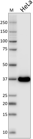
Total cell lysate from HeLa cells (lane 1, 15 µg), NIN3T3 cells (lane 2, 15 µg), U87-MG (lane 3, 15 µg), mouse brain (lane 4, 15 µg) and rat brain (lane 5, 15 µg) were resolved by electrophoresis (4-20% Tris-Glycine gel), transferred to nitrocellulose, and probed with purified anti-β actin antibody (clone 2F1-1). Proteins were visualized using an HRP Goat anti-mouse IgG Antibody and chemiluminescence detection. -

Day-three cultured postnatal C57BL/6 mouse brain cells were fixed with 1% paraformaldehyde (PFA) for 10 minutes, permeabilized with 0.5% Triton X-100 for 10 minutes, and blocked with 5% FBS for 30 minutes. Then the cells were intracellularly stained with 2.5 µg/ml of purified anti- β-actin (clone 2F1-1) in blocking buffer, overnight at 4°C, followed by Alexa Fluor® 594 anti-mouse IgG (red) staining for 2 hours at 4°C. Nuclei were counterstained with DAPI (blue). The image was captured with 40X objective. -

IHC staining using purified anti-β-actin antibody (clone 2F1-1) on formalin-fixed paraffin-embedded human kidney tissue. The tissue was incubated with 5 μg/mL of anti-β-actin antibody overnight at 4°C, followed by incubation with 2.5 μg/mL of Alexa Fluor® 647 goat anti-mouse IgG antibody (red) (Cat. No. 405322) for one hour at room temperature. Nuclei were counterstained with DAPI (blue) (Cat. No. 422801), and the slide was mounted with ProLong™ Gold Antifade Mountant. The image was captured with a 40x objective. Scalebar = 50 μM.
| Cat # | Size | Price | Quantity Check Availability | Save | ||
|---|---|---|---|---|---|---|
| 643801 | 25 µg | £70 | ||||
| 643802 | 100 µg | £157 | ||||
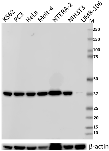
β-actin is a ubiquitously expressed and highly conserved 42 kD cytoplasmic protein involved in cell motility. This critical cytoskeletal component can be disrupted by drugs such as cytochalasin. Because β-actin is ubiquitously expressed in all eukaryotic cells, it is frequently used as a loading control for assays involving protein detection (such as Western blotting).
Product DetailsProduct Details
- Verified Reactivity
- Human, Mouse, Rat
- Antibody Type
- Monoclonal
- Host Species
- Mouse
- Formulation
- Phosphate-buffered solution, pH 7.2, containing 0.09% sodium azide.
- Preparation
- The antibody was purified by affinity chromatography.
- Concentration
- 0.5 mg/ml
- Storage & Handling
- The antibody solution should be stored undiluted between 2°C and 8°C.
- Application
-
WB - Quality tested
ICC, IHC-P - Verified - Recommended Usage
-
Each lot of this antibody is quality control tested by Western blotting. Western blotting, suggested working dilution(s): Use 10 µL per 5 mL antibody dilution buffer for each mini-gel. For immunocytochemistry, a concentration range of 1.0 - 5.0 µg/mL is recommended. For immunohistochemistry, a concentration range of 5 - 10 µg/mL is suggested. It is recommended that the reagent be titrated for optimal performance for each application.
- Application Notes
-
The binding of this antibody to its target is sensitive to salt concentration. For consistent results, please use TBS/T buffer for Western blotting that contains 0.15 M NaCl as indicated in the BioLegend recommended protocol.
-
Application References
(PubMed link indicates BioLegend citation) -
- Lawson BR, et al. 2007. J. Immunol. 178:5366.
- Joyce CW, et al. 2006. J. Biol. Chem. 281:33053.
- Yanagiya T,et al.2007.Obesity.15:572.
- KishidaT,et al. 2007.J. Immunol. 179:8554.
- Ouimet M, et al. 2008. Arterisocler Thromb Vasc Biol. 28:1144.
- Toltlt LJ,et al. 2008.J. Immunol. 181:2165.
- Sawada. T,et al. 2008. J. Biol. Chem. 283:26820.
- Ikeda D, et al. 2008. Endocrinology. 149:6037.
- Ikeda Y, et al. 2010. Mol Endocrinol. 24:1338. PubMed
- Kaieda S, et al. 2010. J. Biol Chem. 285:21478. PubMed
- Cambos M, et al. 2011. J. Leukoc. Biol. 89:157. PubMed
- Product Citations
-
- RRID
-
AB_2223201 (BioLegend Cat. No. 643801)
AB_2223199 (BioLegend Cat. No. 643802)
Antigen Details
- Structure
- Actin, cytoplasmic 1
- Distribution
-
Ubiquitously expressed in the cytoplasm of all eukaryotic cells
- Function
- Actins are highly conserved proteins that are involved in cell motility
- Biology Area
- Cell Biology, Neuroscience, Neuroscience Cell Markers
- Molecular Family
- Microfilaments
- Antigen References
-
1. Hanukoglu I, et al. 1983. J. Mol. Biol. 163:673.
2. Nakajima-Iijima S, et al. 1985. Proc. Natl. Acad. Sci. 82:6133.
3. Ponte P, et al. 1984. Nucleic Acids Res. 12:1687. - Gene ID
- 11461 View all products for this Gene ID
- UniProt
- View information about beta-actin on UniProt.org

Related FAQs
Other Formats
View All β-Actin Reagents Request Custom Conjugation| Description | Clone | Applications |
|---|---|---|
| Alexa Fluor® 594 anti-β-actin | 2F1-1 | ICC,IHC-P |
| Purified anti-β-actin | 2F1-1 | WB,ICC,IHC-P |
| Brilliant Violet 421™ anti-β-actin | 2F1-1 | ICC |
| Direct-Blot™ HRP anti-β-actin | 2F1-1 | WB |
| Alexa Fluor® 647 anti-β-actin | 2F1-1 | IHC-P |
| Alexa Fluor® 488 anti-β-actin | 2F1-1 | IHC-P |
Customers Also Purchased
Compare Data Across All Formats
This data display is provided for general comparisons between formats.
Your actual data may vary due to variations in samples, target cells, instruments and their settings, staining conditions, and other factors.
If you need assistance with selecting the best format contact our expert technical support team.
-
Alexa Fluor® 594 anti-β-actin

MCF7 mammary gland cells were fixed with cold methanol for t... 
Human paraffin-embedded intestine was prepared with standard... -
Purified anti-β-actin

Total cell lysate from HeLa cells (lane 1, 15 µg), NIN3T3 ce... 
Day-three cultured postnatal C57BL/6 mouse brain cells were ... 
IHC staining using purified anti-β-actin antibody (clone 2F1... -
Brilliant Violet 421™ anti-β-actin

NIT/3T3 cells were fixed with 1% paraformaldehyde (PFA) for ... -
Direct-Blot™ HRP anti-β-actin

Total cell lysate from HeLa cells (lane 1, 15 µg), NIH-3T3 c... -
Alexa Fluor® 647 anti-β-actin

Human paraffin-embedded colon tissue slices were prepared wi... -
Alexa Fluor® 488 anti-β-actin

Human paraffin-embedded colon tissue slices were prepared wi...


Follow Us