 Login / Register


- Clone
- 2E1.E9 (See other available formats)
- Regulatory Status
- RUO
- Other Names
- Glial fibrillary acid protein
- Isotype
- Mouse IgG2b
- Ave. Rating
- Submit a Review
- Product Citations
- publications

-

C57BL/6 mouse frozen brain tissue was fixed with 4% paraformaldehyde (PFA) for ten minutes, permeabilized with 0.5 % Triton X-100 for ten minutes, and blocked with 5% FBS for 30 minutes. Then the tissue was stained with 1.25 µg/ml of Brilliant Violet 421™ anti-GFAP (clone 2E1.E9, blue) overnight at 4°C. Nuclei were counterstained with DRAQ5™ (red). The image was captured with a 10X objective. -

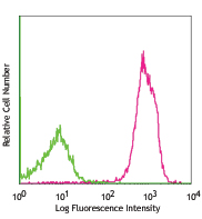
Day-three cultured postnatal C57BL/6 mouse brain cells were fixed with 1% paraformaldehyde (PFA) for ten minutes, permeabilized with 0.5% Triton X-100 for ten minutes, and blocked with 5% FBS for 30 minutes. Then, the cells were intracellularly stained with 5 µg/ml of anti-GFAP (clone 2E1.E9) Brilliant Violet 421™ in 5% FBS overnight at 4°C. Nuclei were counterstained with DRAQ5™ (blue). The image was captured with a 40X objective. -

Human paraffin-embedded human cerebellum tissue slices were prepared with a standard protocol of deparaffinization and rehydration. Antigen retrieval was done with Tris-Buffered Saline 1X (1.0M, pH 7.4) at 95°C for 40 minutes. Tissue was washed with PBS/0.05% Tween 20 twice for five minutes and blocked with 5% FBS and 0.2% gelatin for 30 minutes. Then, the tissue was stained with Brilliant Violet 421™ anti-GFAP Antibody (clone 2E1.E9, blue) at 4°C overnight. The nuclei were counter stained with DRAQ5™ (red). The image was captured with a 10X objective.
| Cat # | Size | Price | Quantity Check Availability | Save | ||
|---|---|---|---|---|---|---|
| 644710 | 100 µg | £298 | ||||
GFAP is expressed exclusively in astrocytes in the central nervous system. The protein is a member of the intermediate filament family of proteins which form networks providing support and strength to cells. This antibody does not cross-react with other intermediate filaments such as vimentin, neurofilament proteins, desmin and others. More than 50 GFAP mutations have been identified to be associated with the Alexander disease.
Product DetailsProduct Details
- Verified Reactivity
- Human, Mouse, Rat
- Antibody Type
- Monoclonal
- Host Species
- Mouse
- Immunogen
- Bovine spinal cord homogenate
- Formulation
- Phosphate-buffered solution, pH 7.2, containing 0.09% sodium azide and BSA (origin USA).
- Preparation
- The antibody was purified by affinity chromatography and conjugated with Brilliant Violet 421™ under optimal conditions.
- Concentration
- 0.2 mg/mL
- Storage & Handling
- The antibody solution should be stored undiluted between 2°C and 8°C, and protected from prolonged exposure to light. Do not freeze.
- Application
-
IHC-F - Quality tested
ICC, IHC-P - Verified - Recommended Usage
-
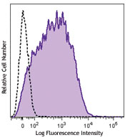
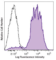
Each lot of this antibody is quality control tested by immunohistochemical staining on frozen tissue sections. For immunohistochemistry, a concentration range of 1.25 - 5.0 µg/mL is suggested. For immunocytochemistry, a concentration range of 2.5 - 5.0 μg/mL is recommended. For immunohistochemical staining on formalin-fixed paraffin-embedded tissue sections, the suggested use of this reagent is 5.0 - 10 µg per mL. It is recommended that the reagent be titrated for optimal performance for each application.
Brilliant Violet 421™ excites at 405 nm and emits at 421 nm. The standard bandpass filter 450/50 nm is recommended for detection. Brilliant Violet 421™ is a trademark of Sirigen Group Ltd.
Learn more about Brilliant Violet™.
This product is subject to proprietary rights of Sirigen Inc. and is made and sold under license from Sirigen Inc. The purchase of this product conveys to the buyer a non-transferable right to use the purchased product for research purposes only. This product may not be resold or incorporated in any manner into another product for resale. Any use for therapeutics or diagnostics is strictly prohibited. This product is covered by U.S. Patent(s), pending patent applications and foreign equivalents. - Excitation Laser
-
Violet Laser (405 nm)
-
Application References
(PubMed link indicates BioLegend citation) -
- McLendon RE and Bigner DD. 1994. Brain Pathol. 4:221.
- Liu W, et al. 2011. Proteomics 11:3556. (FC) PubMed
- Product Citations
-
- RRID
-
AB_2566685 (BioLegend Cat. No. 644710)
Antigen Details
- Structure
- Around 50 kD
- Distribution
-
Only expressed in astrocytes in the central nervous system.
- Function
- Together with other proteins to form intermediate filaments which supports astroglial cells. Astroglial cells support and nourish cells in the brain and spinal cord. If brain cells are injured through trauma or disease, astroglial cells react by rapidly prodcing more GFAP protein.
- Cell Type
- Astrocytes
- Biology Area
- Cell Biology, Neuroscience, Neuroscience Cell Markers
- Molecular Family
- Intermediate Filaments
- Gene ID
- 2670 View all products for this Gene ID
- UniProt
- View information about GFAP on UniProt.org
Related Pages & Pathways
Pathways


Related FAQs
- What is the F/P ratio range of our BV421™ format antibody reagents?
-
It is lot-specific. On average it ranges between 2-4.
Other Formats
View All GFAP Reagents Request Custom Conjugation| Description | Clone | Applications |
|---|---|---|
| Purified anti-GFAP | 2E1.E9 | WB,ICC,IHC-F,IHC-P,FC |
| Alexa Fluor® 488 anti-GFAP | 2E1.E9 | IHC-F,ICC,IHC-P,SB |
| Alexa Fluor® 647 anti-GFAP | 2E1.E9 | IHC-F,IHC-P,SB |
| Alexa Fluor® 594 anti-GFAP | 2E1.E9 | IHC-F,IHC-P,SB |
| Brilliant Violet 421™ anti-GFAP | 2E1.E9 | IHC-F,ICC,IHC-P |
Customers Also Purchased
Compare Data Across All Formats
This data display is provided for general comparisons between formats.
Your actual data may vary due to variations in samples, target cells, instruments and their settings, staining conditions, and other factors.
If you need assistance with selecting the best format contact our expert technical support team.
-
Purified anti-GFAP

Total cell lysate from HeLa (negative control), U87-MG and t... 
U251 cells were fixed with 4% paraformaldehyde (PFA) for ten... 
C57BL/6 mouse frozen brain section was fixed with 4% parafor... 
IHC staining of Purified anti-GFAP (clone 2E1.E9) on formali... 
IHC staining of purified anti-GFAP (clone 2E1.E9) on formali... -
Alexa Fluor® 488 anti-GFAP

C57BL/6 mouse frozen brain section was fixed with 4% parafor... 
Day-three cultured postnatal C57BL/6 mouse brain cells were ... 
IHC staining of Alexa Fluor® 488 anti-GFAP (clone 2E1.E9) on... -
Alexa Fluor® 647 anti-GFAP

C57BL/6 mouse frozen cerebellum section was fixed with 4% pa... 
Human paraffin-embedded cerebellum tissue slices were prepar... 
Human paraffin-embedded cerebellum tissue slices were prepar... -
Alexa Fluor® 594 anti-GFAP

C57BL/6 mouse frozen cerebellum section was fixed with 4% pa... 
IHC staining of Alexa Fluor® 594 anti-GFAP (clone 2E1.E9) on... -
Brilliant Violet 421™ anti-GFAP

Day-three cultured postnatal C57BL/6 mouse brain cells were ... 
C57BL/6 mouse frozen brain tissue was fixed with 4% paraform... 
Human paraffin-embedded human cerebellum tissue slices were ...


Follow Us