 Login / Register


- Clone
- C7 (See other available formats)
- Regulatory Status
- RUO
- Other Names
- CD314
- Isotype
- Armenian Hamster IgG
- Ave. Rating
- Submit a Review
- Product Citations
- publications

-

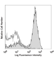
C57BL/6 mouse splenocytes were stained with NK1.1 APC (clone PK136) and biotin NKG2D (clone C7, left) or Armenian Hamster IgG isotype control (clone HTK888, right) followed by SAV-PE.
| Cat # | Size | Price | Quantity Check Availability | Save | ||
|---|---|---|---|---|---|---|
| 115703 | 50 µg | £65 | ||||
NKG2D is a lectin-like type II transmembrane protein also known as CD314. It is expressed on NK cells, a subset of CD8+ T cells, γ/δ T cells and NK1.1+ T cells, as well as in vitro induced LAK cells. NKG2D serves as a stimulatory immunoreceptor to activate NK cells via the non-covalently associated DAP10 or DAP12 adaptor. Several molecules have been identified as the ligands for NKG2D, including minor histocompatibility molecule, H60, UL16-binding protein-like transcript 1 (Mult1, and a family of retinoic acid early transcript 1 (Rae1) in mice, MHC class-I chain-related protein A (MICA), MICB, and UL16-binding proteins (ULBPs) in humans. present in both mice and humans. NKG2D ligands trigger cytokine (IFN-γ, GM-CSF, TNF-α, MIP1β and others) and granzyme release from NK cells. The C7 antibody has been reported to block NK cell killing of targets expressing NKG2D ligands and induce redirected lysis.
Product DetailsProduct Details
- Verified Reactivity
- Mouse
- Antibody Type
- Monoclonal
- Host Species
- Armenian Hamster
- Immunogen
- Soluble mouse NKG2D
- Formulation
- Phosphate-buffered solution, pH 7.2, containing 0.09% sodium azide.
- Preparation
- The antibody was purified by affinity chromatography, and conjugated with biotin under optimal conditions.
- Concentration
- 0.5 mg/ml
- Storage & Handling
- The antibody solution should be stored undiluted between 2°C and 8°C. Do not freeze.
- Application
-
FC - Quality tested
- Recommended Usage
-
Each lot of this antibody is quality control tested by immunofluorescent staining with flow cytometric analysis. For flow cytometric staining, the suggested use of this reagent is ≤ 0.5 µg per 106 cells in 100 µl volume. It is recommended that the reagent be titrated for optimal performance for each application.
- Application Notes
-
Additional reported (for the relevant formats) applications include: blocking1,2,4,5 and inducing redirected cell lysis1,2. To reduce non-specific binding to cells bearing Fc-receptors, pre-incubation of cells with anti-mouse CD16/CD32, clone 93 (Cat. No. 101301/101302), is recommended prior to immunofluorescent staining. For most successful immunofluorescent staining results, it may be important to maximize signal over background by using a relatively bright fluorochrome-antibody conjugate (Cat. No. 115706) or by using a high sensitivity, three-layer staining technique (e.g., including a biotinylated antibody (Cat. No. 115704) or biotinylated anti-Armenian hamster IgG (Cat. No. 405501) second step, followed by SAv-PE (Cat. No. 405204)). The Ultra-LEAF™ purified antibody (Endotoxin <0.1 EU/µg, Azide-Free, 0.2 µm filtered) is recommended for functional assays (Cat. No. 115713 & 115714).
-
Application References
(PubMed link indicates BioLegend citation) -
- Gilfillan S, et al. 2002. Nat. Immunol. 3:1150. (Block)
- Ho EL, et al. 2002. J. Immunol. 169:3667. (Block)
- Maeda M, et al. 2004. J. Immunol. 172:6115. (FC)
- Andoniou CE, et al. 2005. Nature Immunology 6:1011. (Block)
- O'Leary JG, et al. 2006. Nature Immunology 7:507. (Block)
- Tang X, et al. 2006. J. Immunol. 177:7645. (FC)
- Product Citations
-
- RRID
-
AB_313664 (BioLegend Cat. No. 115703)
Antigen Details
- Structure
- C-type lectin
- Distribution
-
NK and NK-T cells
- Function
- NK cytolytic killing of target cells expressing NKG2D ligands, costimulation NK cells
- Ligand/Receptor
- Minor histocompatibility molecule H60, retinoic acid early inducible 1 (RAE-1) family
- Cell Type
- NK cells, NKT cells
- Biology Area
- Costimulatory Molecules, Immunology
- Molecular Family
- CD Molecules
- Antigen References
-
1. Vance RE, et al. 1999. J. Exp. Med. 190:1801.
2. Vance RE, et al. 1998. J. Exp. Med. 188:1841.
3. Lohwasser S, et al. 1999. Eur. J. Immunol. 29:755.
4. Yokoyama WM. 1998. Curr. Opin. Immunol. 10:298.
5. Gilfillan S, et al. 2002. Nat. Immunol. 3:1150.
6. Ho EL, et al. 2002. J. Immunol. 169:3667. - Gene ID
- 27007 View all products for this Gene ID
- UniProt
- View information about CD314 on UniProt.org
Related Pages & Pathways
Pathways


Related FAQs
- How many biotin molecules are per antibody structure?
- We don't routinely measure the number of biotins with our antibody products but the number of biotin molecules range from 3-6 molecules per antibody.
Other Formats
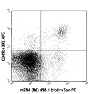
View All CD314 Reagents Request Custom Conjugation| Description | Clone | Applications |
|---|---|---|
| Biotin anti-mouse CD314 (NKG2D) | C7 | FC |
| PE anti-mouse CD314 (NKG2D) | C7 | FC |
| FITC anti-mouse CD314 (NKG2D) | C7 | FC |
| Ultra-LEAF™ Purified anti-mouse CD314 (NKG2D) | C7 | FC,Block |
Customers Also Purchased
Compare Data Across All Formats
This data display is provided for general comparisons between formats.
Your actual data may vary due to variations in samples, target cells, instruments and their settings, staining conditions, and other factors.
If you need assistance with selecting the best format contact our expert technical support team.
-
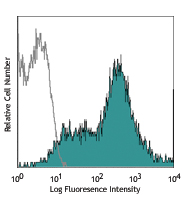
Biotin anti-mouse CD314 (NKG2D)

C57BL/6 mouse splenocytes were stained with NK1.1 APC (clone... -
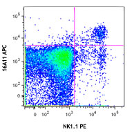
PE anti-mouse CD314 (NKG2D)

C57BL/6 splenocytes stained with C7 PE and NK1.1 FITC -
FITC anti-mouse CD314 (NKG2D)

C57BL/6 mouse splenocytes were stained with NK1.1 APC (clone... -
Ultra-LEAF™ Purified anti-mouse CD314 (NKG2D)

C57BL/6 mouse splenocytes were stained with NK1.1 APC (clone...


Follow Us