 Login / Register


- Clone
- 5B2 (See other available formats)
- Regulatory Status
- RUO
- Other Names
- CEACAM1,5,6,8
- Isotype
- Mouse IgG1, κ
- Ave. Rating
- Submit a Review
- Product Citations
- publications

-

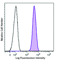
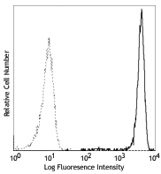
Human colorectal adenocarcinoma cell line were stained with CD66a/b/c/e (clone 5B2) Alexa Fluor® 647 (filled histogram) or mouse IgG1, κ (clone MOPC-21) Alexa Fluor® 647 isotype control (open histogram). -

Human paraffin-embedded colon tissue slices were prepared with a standard protocol of deparaffinization and rehydration. Antigen retrieval was done with Citrate buffer 1X pH 6.0 at 95°C for 40 minutes. Tissue was washed with PBS/0.05% Tween 20 twice for five minutes and blocked with 5% FBS and 0.2% gelatin for 30 minutes. Then, the tissue was stained with 10 µg/mL of anti-human CD66a/b/c/e (clone 5B2) Alexa Fluor® 647 (red) and anti-Vimentin (clone O91D3) Alexa Fluor® 594 (green) at 4°C overnight. Nuclei were counterstained with DAPI (blue). The image was scanned with a 10X objective and stitched with the MetaMorph® software. -

Human paraffin-embedded spleen tissue slices were prepared with a standard protocol of deparaffinization and rehydration. Antigen retrieval was done with Citrate buffer 1X pH 6.0 at 95°C for 40 minutes. Tissue was washed with PBS/0.05% Tween 20 twice for five minutes and blocked with 5% FBS and 0.2% gelatin for 30 minutes. Then, the tissue was stained with 10 µg/mL of anti-human CD66a/b/c/e (clone 5B2) Alexa Fluor® 647 (red) at 4°C overnight. Nuclei were counterstained with DAPI (blue). The image was scanned with a 10X objective and stitched with Metamorph® software.
| Cat # | Size | Price | Quantity Check Availability | Save | ||
|---|---|---|---|---|---|---|
| 398905 | 25 tests | £97 | ||||
| 398906 | 100 tests | £233 | ||||
The 5B2 monoclonal antibody reacts with the antigen epitopes shared by CD66a, b, c and e, which are members of the CEA (carcinoembryonic antigen) family of the Ig superfamily. CD66 plays a role in hemophilic and heterophilic adhesion. CD66a, also known as CEACAM1 and BGP (biliary glycoprotein), is mainly expressed on granulocytes, binds to CD66a, c, e, and CD62E. CD66c (known as CEACAM6, NCA) is expressed on granulocytes and epithelial cells. The ligands of CD66c are CD66a-e, CD62E and Galectins. CD66d is a major granulocyte receptor mediating recognition and efficient opsonin-independent phagocytosis of CEACAM-binding microorganisms, including Neisseria, Moraxella and Haemophilus species, thus playing an important role in the clearance of pathogens by the innate immune system. CD66e molecule is also called CEA or CEACAM5, and is primarily found on epithelial cells. CD66e binds to CD66a, c and e.
Product DetailsProduct Details
- Verified Reactivity
- Human
- Antibody Type
- Monoclonal
- Host Species
- Mouse
- Formulation
- Phosphate-buffered solution, pH 7.2, containing 0.09% sodium azide and BSA (origin USA)
- Preparation
- The antibody was purified by affinity chromatography and conjugated with Alexa Fluor® 647 under optimal conditions.
- Concentration
- Lot-specific (to obtain lot-specific concentration and expiration, please enter the lot number in our Certificate of Analysis online tool.)
- Storage & Handling
- The antibody solution should be stored undiluted between 2°C and 8°C, and protected from prolonged exposure to light. Do not freeze.
- Application
-
FC - Quality tested
IHC-P - Verified - Recommended Usage
-
Each lot of this antibody is quality control tested by immunofluorescent staining with flow cytometric analysis. For flow cytometric staining, the suggested use of this reagent is 5 µL per million cells in 100 µL staining volume or 5 µL per 100 µL of whole blood. For immunohistochemistry, a concentration range of 5.0 - 10.0 µg/mL is suggested. It is recommended that the reagent be titrated for optimal performance for each application.
* Alexa Fluor® 647 has a maximum emission of 668 nm when it is excited at 633 nm / 635 nm.
Alexa Fluor® and Pacific Blue™ are trademarks of Life Technologies Corporation.
View full statement regarding label licenses - Excitation Laser
-
Red Laser (633 nm)
- RRID
-
AB_2888720 (BioLegend Cat. No. 398905)
AB_2888720 (BioLegend Cat. No. 398906)
Antigen Details
- Structure
- Ig superfamily, CEA antigen group, GPI-linked glycoprotein
- Distribution
-
Granulocytes, epithelial cells
- Function
- Homophilic and heterophilic adhesion
- Ligand/Receptor
- CD66a-e, CD62E, galectins
- Cell Type
- Epithelial cells, Granulocytes
- Biology Area
- Immunology
- Molecular Family
- Adhesion Molecules, CD Molecules
- Antigen References
-
- Zola H, et al. 2007. Leukocyte and Stromal Cell Molecules:The CD Markers. Wiley-Liss. A John Wiley & Sons Inc. Publication
- Chen T, et al. 2001. J Biol Chem. 276:17413.
- Wang Z, et al. 2016. Mol Clin Oncol. 5:731.
- Swords DS, et al. 2016. Onco Targets Ther. 9:7459
- Gene ID
- 1088 View all products for this Gene ID 634 View all products for this Gene ID 4680 View all products for this Gene ID 1048 View all products for this Gene ID
- UniProt
- View information about CD66a/b/c/e on UniProt.org


Related FAQs
Other Formats
View All CD66a/b/c/e Reagents Request Custom Conjugation| Description | Clone | Applications |
|---|---|---|
| Purified anti-human CD66a/b/c/e | 5B2 | IHC-P,FC |
| Alexa Fluor® 647 anti-human CD66a/b/c/e Antibody | 5B2 | FC,IHC-P |
Customers Also Purchased
Compare Data Across All Formats
This data display is provided for general comparisons between formats.
Your actual data may vary due to variations in samples, target cells, instruments and their settings, staining conditions, and other factors.
If you need assistance with selecting the best format contact our expert technical support team.
-
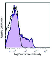
Purified anti-human CD66a/b/c/e

Human paraffin-embedded colon tissue slices were prepared wi... 
Human paraffin-embedded spleen tissue slices were prepared w... 
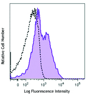
Human colorectal adenocarcinoma cell line HT-29 stained with... -
Alexa Fluor® 647 anti-human CD66a/b/c/e Antibody

Human colorectal adenocarcinoma cell line were stained with ... 
Human paraffin-embedded colon tissue slices were prepared wi... 
Human paraffin-embedded spleen tissue slices were prepared w...


Follow Us