 Login / Register


- Clone
- MI15 (See other available formats)
- Regulatory Status
- RUO
- Workshop
- HCDM listed
- Other Names
- B-B4
- Isotype
- Mouse IgG1, κ
- Ave. Rating
- Submit a Review
- Product Citations
- publications

-

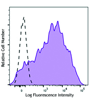
Human myeloma cell line U266 was stained with CD138 (clone MI15) Alexa Fluor® 647 (filled histogram) or mouse IgG1, κ Alexa Fluor® 647 isotype control (open histogram). -

Confocal image of human skin sample acquired using the IBEX method of highly multiplexed antibody-based imaging: CD117 (magenta) in Cycle 3, CD138 (cyan) in Cycle 4, and Vimentin (blue) in Cycle 5. Tissues were prepared using ~1% (vol/vol) formaldehyde and a detergent. Following fixation, samples are immersed in 30% (wt/vol) sucrose for cryoprotection. Images are courtesy of Drs. Andrea J. Radtke and Ronald N. Germain of the Center for Advanced Tissue Imaging (CAT-I) in the National Institute of Allergy and Infectious Diseases (NIAID, NIH). -

Confocal image of human metastatic lymph node sample acquired using the IBEX method of highly multiplexed antibody-based imaging: CD8 (green) in Cycle 1 and CD138 (blue) in Cycle 5. Tissues were prepared using ~1% (vol/vol) formaldehyde and a detergent. Following fixation, samples are immersed in 30% (wt/vol) sucrose for cryoprotection. Images are courtesy of Drs. Andrea J. Radtke and Ronald N. Germain of the Center for Advanced Tissue Imaging (CAT-I) in the National Institute of Allergy and Infectious Diseases (NIAID, NIH).
| Cat # | Size | Price | Quantity Check Availability | Save | ||
|---|---|---|---|---|---|---|
| 356523 | 25 tests | £89 | ||||
| 356524 | 100 tests | £238 | ||||
CD138, a member of the syndecan protein family, is a type I integral membrane heparin sulfate proteoglycan also known as Syndecan-1. Syndecan-1 participates in cell proliferation, cell migration, and cell-matrix adhesion via interaction with collagen, fibronectin, and other soluble molecules (such as FGF-basic). It is expressed on normal and malignant human plasma cells, pre-B cells, epithelial cells, and endothelial cells, but not on mature circulating B-lymphocytes. It is also expressed on some non-hematopoietic cells, including embryonic mesenchymal cells, vascular smooth muscle cells, endothelial cells, and neural cells.
Product DetailsProduct Details
- Verified Reactivity
- Human
- Antibody Type
- Monoclonal
- Host Species
- Mouse
- Immunogen
- A mixture of U266 and XG-1 human myeloma cell lines.
- Formulation
- Phosphate-buffered solution, pH 7.2, containing 0.09% sodium azide and BSA (origin USA)
- Preparation
- The antibody was purified by affinity chromatography and conjugated with Alexa Fluor® 647 under optimal conditions.
- Concentration
- Lot-specific (to obtain lot-specific concentration and expiration, please enter the lot number in our Certificate of Analysis online tool.)
- Storage & Handling
- The antibody solution should be stored undiluted between 2°C and 8°C, and protected from prolonged exposure to light. Do not freeze.
- Application
-
FC - Quality tested
SB - Community verified
SB - Reported in the literature, not verified in house - Recommended Usage
-
Each lot of this antibody is quality control tested by immunofluorescent staining with flow cytometric analysis. For flow cytometric staining, the suggested use of this reagent is 5 µl per million cells in 100 µl staining volume or 5 µl per 100 µl of whole blood.
* Alexa Fluor® 647 has a maximum emission of 668 nm when it is excited at 633 nm / 635 nm.
Alexa Fluor® and Pacific Blue™ are trademarks of Life Technologies Corporation.
View full statement regarding label licenses - Excitation Laser
-
Red Laser (633 nm)
- Application Notes
-
The epitope recognized by MI15 is found within the ectodomain of the CD138 core protein. It has been reported that MI15 blocks the binding of clone B-B4 but not clone DL-101 by flow cytometric analysis. Clones DL-101 and MI15 exhibit differential staining patterns on in vitro generated plasma cells and some CD138+ cell lines.4
Additional reported applications for the relevant formats include: immunofluorescent staining1, Western blotting2, immunohistochemical staining of formalin-fixed paraffin-embedded frozen tissue sections3, and spatial biology (IBEX)5,6. - Additional Product Notes
-
For use in spatial biology, this antibody has been demonstrated for use in immunohistochemistry using IBEX (Reported in the literature, not verified in house) and the NanoString GeoMx® Digital Spatial Profiler.
IBEX: Iterative Bleaching Extended multi-pleXity (IBEX) is a fluorescent imaging technique capable of highly-multiplexed spatial analysis. The method relies on cyclical bleaching of panels of fluorescent antibodies in order to image and analyze many markers over multiple cycles of staining, imaging, and, bleaching. It is a community-developed open-access method developed by the Center for Advanced Tissue Imaging (CAT-I) in the National Institute of Allergy and Infectious Diseases (NIAID, NIH).
NanoString GeoMx®: This product has been verified for IHC-P (Immunohistochemistry - formalin-fixed paraffin-embedded tissues) on the NanoString GeoMx® Digital Spatial Profiler. The GeoMx® enables researchers to perform spatial analysis of protein and RNA targets in FFPE and fresh frozen human and mouse samples. For more information about our spatial biology products and the GeoMx® platform, please visit our spatial biology page. -
Application References
(PubMed link indicates BioLegend citation) -
- Costes V, et al. 1999. Hum. Pathol. 30:1405. (IF)
- Gattei V, et al. 1999. Br. J. Haematol. 104:152. (WB)
- Bologna-Molina R, et al. 2008. Oral Oncol. 44:805. (IHC)
- Itoua MR, et al. 2014. Biomed. Res. Int. 2014:536482.
- Radtke AJ, et al. 2020. Proc Natl Acad Sci USA. 117:33455-33465. (SB) PubMed
- Radtke AJ, et al. 2022. Nat Protoc. 17:378-401. (SB) PubMed
- Product Citations
-
- RRID
-
AB_2564250 (BioLegend Cat. No. 356523)
AB_2564251 (BioLegend Cat. No. 356524)
Antigen Details
- Structure
- 100-200 kD type I integral transmembrane glycoprotein
- Distribution
-
Plasma cells, pre-B cells, epithelial cells, endothelial cells
- Function
- Adhesion, controls cell morphology, regulates cell growth
- Ligand/Receptor
- FGFb, collagen, fibronectin
- Cell Type
- B cells, Endothelial cells, Epithelial cells, Plasma cells
- Biology Area
- Cell Adhesion, Cell Biology, Cell Motility/Cytoskeleton/Structure, Immunology, Neuroscience, Synaptic Biology
- Molecular Family
- Adhesion Molecules, CD Molecules
- Antigen References
-
1. Sanderson RD, et al. 1992. Cell. Regul. 1:27.
2. Zola H, et al. 2007. Leukocyte and Stromal Cell Molecules: The CD Markers. Wiley-Liss A John Wiley & Sons Inc, Publication. - Gene ID
- 6382 View all products for this Gene ID
- UniProt
- View information about CD138 on UniProt.org
Related Pages & Pathways
Pathways


Related FAQs
- If an antibody clone has been previously successfully used in IBEX in one fluorescent format, will other antibody formats work as well?
-
It’s likely that other fluorophore conjugates to the same antibody clone will also be compatible with IBEX using the same sample fixation procedure. Ultimately a directly conjugated antibody’s utility in fluorescent imaging and IBEX may be specific to the sample and microscope being used in the experiment. Some antibody clone conjugates may perform better than others due to performance differences in non-specific binding, fluorophore brightness, and other biochemical properties unique to that conjugate.
- Will antibodies my lab is already using for fluorescent or chromogenic IHC work in IBEX?
-
Fundamentally, IBEX as a technique that works much in the same way as single antibody panels or single marker IF/IHC. If you’re already successfully using an antibody clone on a sample of interest, it is likely that clone will have utility in IBEX. It is expected some optimization and testing of different antibody fluorophore conjugates will be required to find a suitable format; however, legacy microscopy techniques like chromogenic IHC on fixed or frozen tissue is an excellent place to start looking for useful antibodies.
- Are other fluorophores compatible with IBEX?
-
Over 18 fluorescent formats have been screened for use in IBEX, however, it is likely that other fluorophores are able to be rapidly bleached in IBEX. If a fluorophore format is already suitable for your imaging platform it can be tested for compatibility in IBEX.
- The same antibody works in one tissue type but not another. What is happening?
-
Differences in tissue properties may impact both the ability of an antibody to bind its target specifically and impact the ability of a specific fluorophore conjugate to overcome the background fluorescent signal in a given tissue. Secondary stains, as well as testing multiple fluorescent conjugates of the same clone, may help to troubleshoot challenging targets or tissues. Using a reference control tissue may also give confidence in the specificity of your staining.
- How can I be sure the staining I’m seeing in my tissue is real?
-
In general, best practices for validating an antibody in traditional chromogenic or fluorescent IHC are applicable to IBEX. Please reference the Nature Methods review on antibody based multiplexed imaging for resources on validating antibodies for IBEX.
Other Formats
View All CD138 Reagents Request Custom ConjugationCustomers Also Purchased
Compare Data Across All Formats
This data display is provided for general comparisons between formats.
Your actual data may vary due to variations in samples, target cells, instruments and their settings, staining conditions, and other factors.
If you need assistance with selecting the best format contact our expert technical support team.
-
PE anti-human CD138 (Syndecan-1)

Human myeloma cell line U266 was stained with CD138 (clone M... 
Confocal image of human lymph node sample acquired using the... 
Confocal image of human liver sample acquired using the IBEX... 
Confocal image of human jejunum sample acquired using the IB... -
Purified anti-human CD138 (Syndecan-1)

Human myeloma cell line U266 was stained with purified CD138... 
SeqIF™ (sequential immunofluorescence) staining on COMET™ of... -
APC anti-human CD138 (Syndecan-1)

Human myeloma cell line U266 was stained with CD138 (clone M... -
FITC anti-human CD138 (Syndecan-1)

Human myeloma cell line U266 was stained with CD138 (clone M... -
PerCP/Cyanine5.5 anti-human CD138 (Syndecan-1)

Human myeloma cell line U266 was stained with CD138 (Syndeca... -
Alexa Fluor® 700 anti-human CD138 (Syndecan-1)

Human myeloma cell line U266 was stained with CD138 (clone M... -
PE/Cyanine7 anti-human CD138 (Syndecan-1)

Human myeloma cell line U266 was stained with CD138 (clone M... -
Brilliant Violet 421™ anti-human CD138 (Syndecan-1)

Human myeloma cell line U266 was stained with CD138 (clone M... -
Brilliant Violet 510™ anti-human CD138 (Syndecan-1)

Human myeloma cell line U266 was stained with CD138 (clone M... -
Brilliant Violet 605™ anti-human CD138 (Syndecan-1)

Human myeloma cell line U266 was stained with CD138 (clone M... -
Brilliant Violet 711™ anti-human CD138 (Syndecan-1)

Human myeloma cell line U266 was stained with CD138 (clone M... -
Alexa Fluor® 647 anti-human CD138 (Syndecan-1)

Human myeloma cell line U266 was stained with CD138 (clone M... 
Confocal image of human skin sample acquired using the IBEX ... 
Confocal image of human metastatic lymph node sample acquire... -
Alexa Fluor® 594 anti-human CD138 (Syndecan-1)

U266B1 cells were fixed with 2% paraformaldehyde (PFA) for t... -
PE/Dazzle™ 594 anti-human CD138 (Syndecan-1)

Human myeloma cell line U266 was stained with CD138 (clone M... -
APC/Cyanine7 anti-human CD138 (Syndecan-1)

Human myeloma cell line U266 was stained with CD138 (clone M... -
Pacific Blue™ anti-human CD138 (Syndecan-1)

Human myeloma cell line U266 was stained with Pacific Blue™ ... -
TotalSeq™-A0055 anti-human CD138 (Syndecan-1)
-
Brilliant Violet 785™ anti-human CD138 (Syndecan-1)

Human myeloma cell line U266 was stained with CD138 (clone M... -
Biotin anti-human CD138 (Syndecan-1)

Human myeloma cell line U266 was stained with CD138 (Syndeca... -
TotalSeq™-C0055 anti-human CD138 (Syndecan-1)
-
APC/Fire™ 750 anti-human CD138 (Syndecan-1)

Human myeloma cell line U266 was stained with CD138 (Syndeca... -
TotalSeq™-B0055 anti-human CD138 (Syndecan-1)
-
PE/Cyanine5 anti-human CD138 (Syndecan-1)

U266 (human myeloma cell line) cells were stained with anti-... -
TotalSeq™-D0055 anti-human CD138 (Syndecan-1)
-
PE/Fire™ 640 anti-human CD138 (Syndecan-1)

U266 cells were stained with anti-human CD138 (Syndecan-1) (... -
Spark Violet™ 500 anti-human CD138 (Syndecan-1)

Human myeloma cell line U266 was stained with anti-human CD1... -
FITC anti-human CD138

Typical results from human myeloma cell line U266 stained ei... -
PE/Fire™ 700 anti-human CD138 (Syndecan-1)

U266 cells were stained with anti-human CD138 (Syndecan-1) (... -
Spark Violet™ 423 anti-human CD138 (Syndecan-1)

Human myeloma cell line U266 was stained with anti-human CD1... -
Spark Red™ 718 anti-human CD138 (Syndecan-1) (Flexi-Fluor™)


Follow Us