At BioLegend, we've spent nearly two decades crafting new fluorophores for flow cytometry. With each new option, researchers and colleagues have come to trust the performance of our products, as evidenced by this CiteAb report indicating we are the most cited company for flow cytometry antibodies.


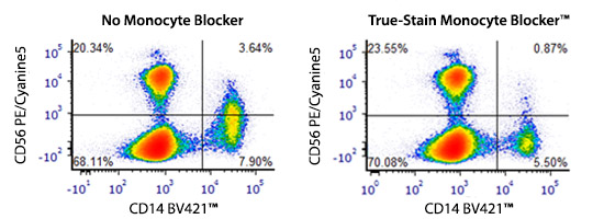
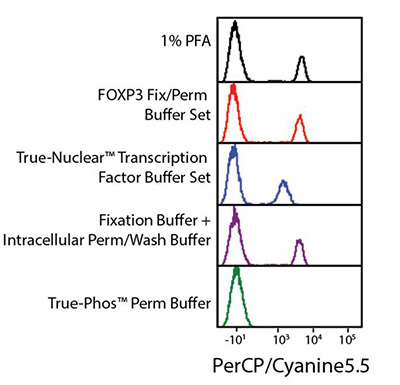
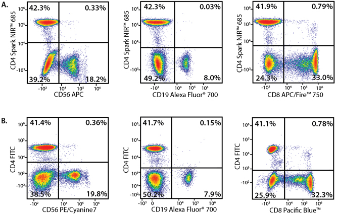
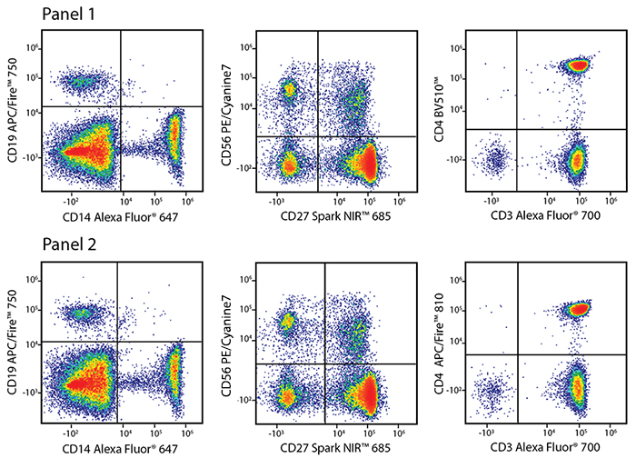
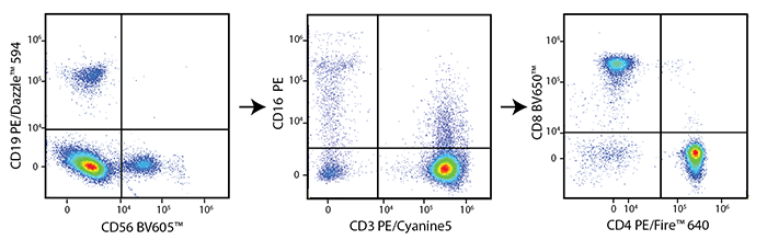
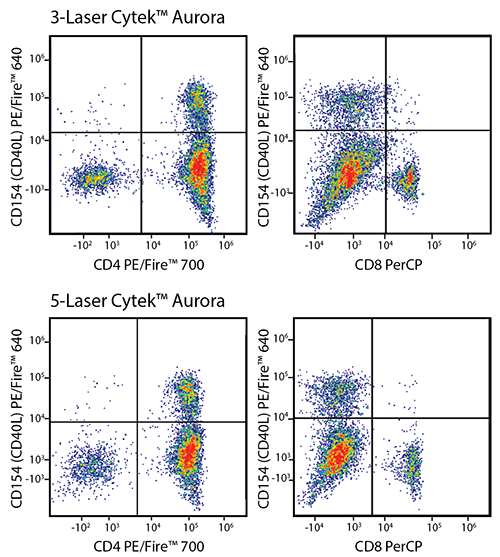
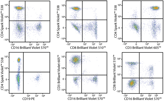
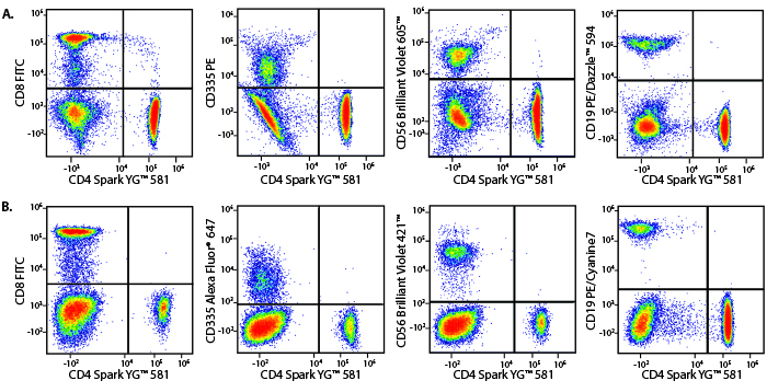
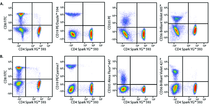
Our diverse array of fluorophores allows you to expand the limits of flow cytometry and develop larger multicolor panels. High level panels (>22 colors) can be built using cytometers capable of spectral detection or the ability to unmix a fluorescent spectrum across an array of emission detectors such as the Cytek™ Aurora or Northern Lights. Some fluorophores, like the Spark Dyes, are specifically designed to be used on these cytometers.


Learn more about the unique advantages offered by each member among the various families of fluorophores and how to best use them in multicolor panels:

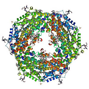
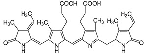
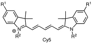
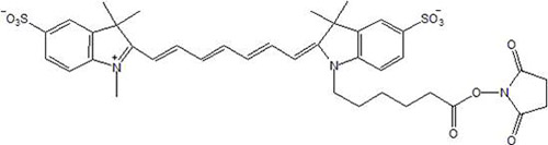
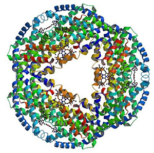
  • Protein-based fluors: PE, APC, PerCP, and their tandems
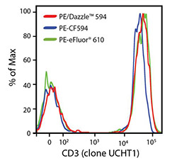
  • Organic fluors: FITC, Alexa Fluor® dyes, Spark Fluors, etc.
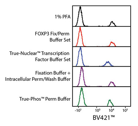
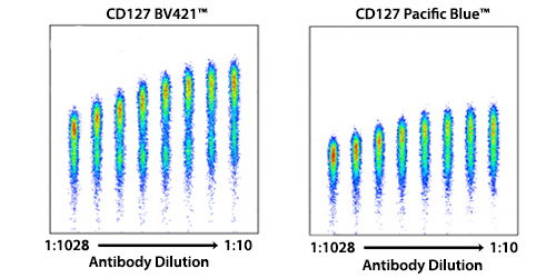
  • Polymers: Brilliant Violet™ Dyes
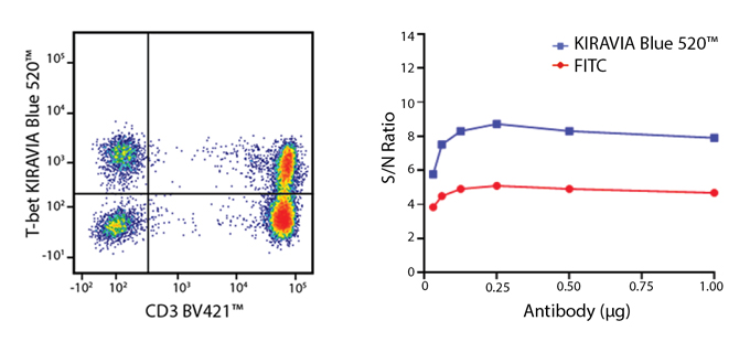
  • KIRAVIA multimers: KIRAVIA Dyes™, a distinct class of multimers with unique fluorescent chemistry.

Two of the most commonly used fluorophores in flow cytometry, R-phycoerythrin (R-PE) and allophycocyanin (APC), are phycobiliproteins originally derived from algae. Both proteins contain fluorescent phycobilin chromophores embedded into a protein scaffolding which accounts for their large size. When phycobiliproteins are exposed to organic solvents like MeOH and EtOH, they denature and are no longer able to fluoresce. Both R-PE and APC are fairly bright fluors and are often used in flow cytometry to detect proteins with low levels of expression.


Another protein-based dye, PerCP, a peridinin-chlorophyll protein complex, is derived from photosynthetic dinoflagellates. Other fluorescent proteins, like eGFP, mCherry, and tdTomato can be fused to a protein of interest to visualize or track cell processes. These are typically derived from sea anemone and jellyfish, and exist as either monomers, dimers, or trimers. All of these protein-based dyes can be used as the donor molecule in FRET pairs. Generally, antibodies conjugated to protein-based flours are not commonly used in standard microscopy as they are more susceptible to photobleaching.

PE


Brightness: 5
Ex Laser: 488 nm, 532 nm, or 561 nm
Ex Max: 565 nm
Em Max: 575 nm
Filter Used: 585/20
Molecular Weight: 240 kDa
Recommended Application(s):
Flow Cytometry

Fluorophore at a Glance:

  • One of the brightest fluorophores and provides high signal-to-noise ratio.
  • A high extinction coefficient and quantum yield make it an ideal donor in a FRET relationship (tandems).
  • Can be detected on almost all cytometers.
  • One of the largest fluorophores,so it may not be optimal for all targets.
  • Though it is more susceptible to photobleaching under continual illumination, PE itself can be used under some high-speed imaging conditions.
  • Sensitive to organic solvent-based fixatives, like those used for phospho flow cytometry staining.
  • All proteins are excited by shorter wavelengths such as UV and Violet lasers. As such,they may have spillover into equivalent channels on those lasers.


View all PE Products.

Structure of R-phycoerythrin


Image from the RCSB PDB (rcsb.org) of PDB ID 1EYX Contreras-Martel, C. et al. 2001. Crystallization and 2.2 A resolution structure of R-phycoerythrin from Gracilaria chilensis:a case of perfect hemihedral twinning. Acta Cystallogr. Sect.D. 57:52-60.

Phycobilin Structure


Structure of phycobilin. R-phycoerythrin is composed of phycobilin chromophores embedded in a protein scaffolding.

Simple organic fluorophores encompass a variety of synthetically-derived dyes. Because of their small size, multiple simple organic fluorophores can be conjugated to a single antibody. Due to their conjugation chemistry, these dyes are flexible and can be easily used to generate custom antibodies. Unlike protein-based fluors, simple organic dyes are not sensitive to organic solvents, like those used in phospho-flow, making them advantageous for these applications. Organic dyes are also soluble, meaning they are less susceptible to aggregation and precipitation. Simple organic dyes are often used in microscopy due to their discrete excitation and emission profiles and are chosen for their limited spillover into other imaging channels. Simple organic dyes are great to stain intracellular or intranuclear targets.

Spark PLUS UV™ 395


Brightness: 4
Ex Laser: 355 nm
Ex Max: 355 nm
Em Max: 385 nm
Filter Used: 379/28
Molecular Weight: 25-30 kDa
Recommended Application(s):
Flow Cytometry

Fluorophore at a Glance:

  • Bright UV-excited alternative to BD Horizon™ BUV395
  • First in a family of brighter Spark Dyes
  • Highly stable to heat and fixatives
  • Can be unmixed from Spark UV™ 387 on spectral cytometers with proper panel design
 

View all Spark PLUS UV™ 395 products.


Learn more about Spark PLUS UV™ 395.

Brilliant Violet™ dyes are organic, light-emitting, diode polymers whose brightness is related to the length of the polymer, but whose spectra remain stable no matter the polymer’s length. These dyes have a high extinction coefficient and quantum yield resulting in high intensity brightness. Brilliant Violet™ dyes are effective over a wide pH range and are compatible with a variety of fixatives.

Brilliant Violet 421™


Brightness: 5
Ex Laser: 405 nm
Ex Max: 405 nm
Em Max: 421 nm
Filter Used: 450/50
Molecular Weight: 60-80 kDa
Recommended Application(s):
Flow Cytometry, Microscopy

Fluorophore at a Glance:

  • A very potent donor with an extinction coefficient of 2,500,000 M-1cm-1 which makes for very potent tandem dyes.
  • In some cases, it can exhibit non-specific binding to itself.
  • Very bright, ideal for detecting low-expressed markers.
 

View all Brilliant Violet 421™ Products.
 

Learn more about Brilliant Violet Dyes.

BV421™ withstands a variety of fixatives

Improved Signal to Noise as compared to Pacific Blue™


Human peripheral blood lymphocytes stained with anti-CD127conjugated to BV421™ and Pacific Blue™ at varying dilutions.

Introducing the KIRAVIA Dyes™, a brand new fluorescent chemistry to advance applications in multicolor flow cytometry and beyond. This class of dyes employs a unique organic backbone that separates fluorophores to minimize quenching effects, thus allowing optimal and higher fluorophore to protein (F:P) ratios, surpassing what is possible with direct conjugations of single fluorophore like FITC.

KIRAVIA Blue 520™


Brightness: 4
Ex Laser: 488 nm
Ex Max: 488 nm
Em Max: 520 nm
Filter Used: 530/30
Molecular Weight: 8 kDa
Recommended Application(s):
Flow Cytometry

Fluorophore at a Glance:

  • An upgrade over FITC in terms of brightness.
  • A bright and spectrally clean fluorophore that only exhibits significant spillover into Spark Blue™ 550. Few, if any, fluors spill into KIRAVIA Blue 520™.
  • Utilize this fluor on ubiquitously expressed markers or markers with varying levels of expression.
 

View all KIRAVIA Blue 520™ Products.
 

Learn more about KIRAVIA Dyes™.

Equivalent fluorophores comparison

Amazing Resolution

Anti-human CD4 (clone SK3), conjugated to FITC (red), Alexa Fluor® 488 (blue), BD Horizon™ Brilliant Blue 515 (green), or KIRAVIA Blue 520™ (purple) was used to stain human lysed whole blood.

(Left) Anti-human CD3 (clone UCHT1) BV421™ and anti-human T-bet (clone 4B10) KIRAVIA Blue 520™ staining on human PBMCs. (Right) Titration curve with FITC or KIRAVIA Blue 520™ conjugated to anti-T-bet antibody.

PE


Brightness: 5
Ex Laser: 488 nm, 532 nm, or 561 nm
Ex Max: 565 nm
Em Max: 575 nm
Filter Used: 585/20
Molecular Weight: 240 kDa
Recommended Application(s):
Flow Cytometry

Fluorophore at a Glance:

  • One of the brightest fluorophores and provides high signal-to-noise ratio.
  • A high extinction coefficient and quantum yield make it an ideal donor in a FRET relationship (tandems).
  • Can be detected on almost all cytometers.
  • One of the largest fluorophores, so it may not be optimal for all targets.
  • Though it is more susceptible to photobleaching under continual illumination, PE itself can be used under some high-speed imaging conditions.
  • Sensitive to organic solvent-based fixatives, like those used for phospho flow cytometry staining.
  • All proteins are excited by shorter wavelengths such as UV and Violet lasers. As such, they may have spillover into equivalent channels on those lasers.


View all PE Products.

Structure of R-phycoerythrin


Image from the RCSB PDB (rcsb.org) of PDB ID 1EYX Contreras-Martel, C. et al. 2001. Crystallization and 2.2 A resolution structure of R-phycoerythrin from Gracilaria chilensis: a case of perfect hemihedral twinning. Acta Cystallogr. Sect.D.57:52-60.

Phycobilin Structure


Structure of phycobilin. R-phycoerythrin is composed of phycobilin chromophores embedded in a protein scaffolding.

PE/Dazzle™ 594


Brightness: 5
Ex Laser: 488 nm, 532 nm, or 561 nm
Ex Max: 565 nm
Em Max: 610 nm
Filter Used: 610/20
Molecular Weight: 245 kDa
Recommended Application(s):
Flow Cytometry

Fluorophore at a Glance:

  • Bright signal with little background.
  • Comparable compensation requirements into PE when compared to spectrally equivalent tandems.
  • Better temperature and photostability as compared to spectrally equivalent tandems.
  • No cross-beam excitation with the red laser.
  • One of the largest fluorophores, so it may not be optimal for all targets.
  • PE/Dazzle™ 594 may exhibit non-specific binding to monocytes and macrophages. Use our True-Stain Monocyte Blocker™ before staining to block this non-specific binding.
 

View all PE/Dazzle™ 594 Products.


Learn more on our PE/Dazzle™ 594 webpage.

Bright Signal and Excellent Resolution


Human peripheral blood lymphocytes stained with anti-human CD3 (clone UCHT1) conjugated to PE/Dazzle™ 594 or other equivalent fluorophores.

True-Stain Monocyte Blocker™ Prevents Non-Specific Binding of PE/Dazzle™ 594 to Monocytes


Human PBMCs were incubated with no monocyte blocker (left) or with 5 µL of True-Stain Monocyte Blocker (right) and then stained with 5 µL/test of the indicated antibodies in 100 µL of cells at 1 x 10⁶cells/mL.

PE/Cyanine5


Brightness: 5
Ex Laser: 488 nm, 532 nm, or 561 nm
Ex Max: 565 nm
Em Max: 670 nm
Filter Used: 660/20
Molecular Weight: 242 kDa
Recommended Application(s):
Flow Cytometry

Fluorophore at a Glance:

  • One of the brightest fluorophores, though this isn’t always an advantage because PE/Cyanine5 heavily spills into a variety of other channels due to significant cross-beam excitation by other lasers,particularly the red laser.   This causes unwanted spillover into the APC or Alexa Fluor® 647 channel and can make population resolution challenging.
  • In a multicolor panel, it may be helpful to use PE/Cyanine5 as the “dump” or exclusion channel to exclude specific cell subsets from analysis.
  • On a spectral unmixing cytometer, PE/Cyanine5 can be distinctly identified from APC and Alexa Fluor® 647, allowing for expansion the number of parameters in a multicolor panel.
  • PE/Cyanine5 may exhibit non-specific binding to monocytes and macrophages. Use our True-Stain Monocyte Blocker™ before staining to block this non-specific binding.
  • One of the largest fluorophores, so it may not be optimal for all targets.
 

View all PE/Cyanine5 Products.

Structure of Cyanine5

True-Stain Monocyte Blocker™ Prevents Non-Specific Binding of PE/Cyanine5 to Monocytes


Human PBMCs were incubated with no monocyte blocker (left) or with 5 µL of True-Stain Monocyte Blocker (right) and then stained with 5 µL/test of the indicated antibodies in 100 µL of cells at 1 x 10⁶cells/mL.

PE/Cyanine7


Brightness: 4
Ex Laser: 488 nm, 532 nm, or 561 nm
Ex Max: 565 nm
Em Max: 774 nm
Filter Used: 780/60
Molecular Weight: 245 kDa
Recommended Application(s):
Flow Cytometry

Fluorophore at a Glance:

  • Tandem dye using PE as the donor and Cyanine7 as the acceptor fluorophore.
  • If a marker may be upregulated, it is useful to put it on PE/Cyanine7 because it is the furthest red-shifted fluorophore excited by the 488/561 nm lasers. So, if expression increases, it won’t affect as many neighboring channels or exacerbate spreading error.
  • One of the largest fluorophores and may not be ideal for intracellular targets.
  • PE/Cyanine7 may exhibit non-specific binding to monocytes and macrophages. Use our True-Stain Monocyte Blocker™ before staining to block this non-specific binding.
 

View all PE/Cyanine7 Products.


Learn more about True-Stain Monocyte Blocker™.


Structure of Cyanine7


PerCP


Brightness: 1
Ex Laser: 488 nm
Ex Max: 482 nm
Em Max: 675 nm
Filter Used: 695/40
Molecular Weight: 35 kDa
Recommended Application(s):
Flow Cytometry

Fluorophore at a Glance:

  • Can have stability issues in response to temperature, fixatives, and light. Consider using PerCP/Cyanine5.5 as a more stable option.
  • Unlike PE which has strong excitation off both the 488 and 561 nm lasers, PerCP has much greater efficiency of excitation off of the 488 nm laser, thus creating a distinct spectral fingerprint from PE.
  • All proteins are excited by shorter wavelengths such as UV and Violet lasers. As such, they may have spillover into equivalent channels on those lasers.
 

View all PerCP Products.

PerCP/Cyanine5.5


Brightness: 2
Ex Laser: 488 nm
Ex Max: 482 nm
Em Max: 690 nm
Filter Used: 695/40
Molecular Weight: 35 kDa
Recommended Application(s):
Flow Cytometry

Fluorophore at a Glance:

  • Tandem dye using PerCP as a donor and Cyanine5.5 as the acceptor fluorophore.
  • Effectively replaces PerCP conjugates in an assay, as Cyanine5.5 stabilizes the environmental sensitivity of PerCP.
  • More photostable alternative when compared to PerCP.
  • While it is more stable, it is still not stable with organic, solvent-based fixatives.
  • PerCP/Cyanine5.5 may exhibit non-specific binding to monocytes and macrophages. Use our True-Stain Monocyte Blocker™ before staining to block this non-specific binding.
 

View all PerCP/Cyanine5.5 Products.


Learn more about True-Stain Monocyte Blocker™.


PerCP/Cyanine5.5 is sensitive to organic solvent-based fixatives


APC


Brightness: 4
Ex Laser: 633 nm or 640 nm
Ex Max: 650 nm
Em Max: 660 nm
Filter Used: 660/20
Molecular Weight: 105 kDa
Recommended Application(s):
Flow Cytometry

Fluorophore at a Glance:

  • A potent donor in a FRET pair.
  • Sensitive to organic solvent-based fixatives, like those used for phospho flow staining.
  • Spectrally distinct from Alexa Fluor® 647 when used on a cytometer capable of spectral unmixing, allowing them to be used together in a single panel. Ideally, these two fluors are not used to detect co-expressed markers.
  • All proteins are excited by shorter wavelengths such as UV and Violet lasers. As such, they may have spillover into equivalent channels on those lasers.
 

View all APC Products.


Structure of Allophycocyanin(APC)


Image from the RCSB PDB (rcsb.org) of PDB ID:2VJT Murray, J.W., Benson, S., Nield, J., Barber, J.The Structure of Allophycocyanin from Gloeobacter Violaceus.


APC/Cyanine7


Brightness: 2
Ex Laser: 633 nm or 640 nm
Ex Max: 650 nm
Em Max: 774 nm
Filter Used: 780/60
Molecular Weight: 109 kDa
Recommended Application(s):
Flow Cytometry

Fluorophore at a Glance:

  • Tandem dye using APC as the donor and Cyanine7 as the acceptor fluorophore.
  • Relatively dim fluorophore, should be used for higher abundance targets.
  • Less stable to temperature, light, and fixative as compared to other spectrally equivalent dyes. Consider using APC/Fire™ 750 as a more stable alternative.
  • If a marker may be upregulated, like an activation or exhaustion marker, it is useful to put it on APC/Cyanine7 because it is the furthest red-shifted fluorophore excited by the 633 nm lasers. So, if expression increases, it won’t affect as many neighboring channels or exacerbate spreading error.
  • APC/Cyanine7 may exhibit non-specific binding to monocytes and macrophages. Use our True-Stain Monocyte Blocker™ before staining to block this non-specific binding.
 

View all APC/Cyanine7 Products.


True-Stain Monocyte Blocker™ Prevents Non-Specific Binding of APC/Cyanine7 to Monocytes


APC/Fire™ 750


Brightness: 2
Ex Laser: 633 nm or 640 nm
Ex Max: 650 nm
Em Max: 774 nm
Filter Used: 780/60
Molecular Weight: 110 kDa
Recommended Application(s):
Flow Cytometry

Fluorophore at a Glance:

  • More temperature and photo-stable alternative for APC/Cyanine7.
  • Improved stability in fixative solutions.
  • Lower compensation requirements than APC/Cyanine7 without compromising brightness.
  • Minimal non-specific binding to monocytes.
  • Like PE/Cyanine7, it is a good fluor to use to detect markers of variable expression because its emission is red-shifted away from neighboring channels.
  • APC/Fire™ 750 may exhibit non-specific binding to monocytes and macrophages. Use our True-Stain Monocyte Blocker™ before staining to block this non-specific binding.
 

View all APC/Fire™ 750 Products.
 

Learn more on our APC/Fire™ 750 webpage.
 

Learn more about True-Stain Monocyte Blocker™.

Comparisons with Spectrally Equivalent Fluors:


Improved Stability in Fixative


Human whole blood was stained for 20 min with CD3 (SK7) conjugates of APC/Fire™ 750, APC/Cyanine7, APC-H7 followed by RBC lysis and wash steps. Cells were then treated with either PBS control, 1%PFA in PBS, 4%PFA followed by.1%saponin, BioLegend's True-Nuclear™ Fix/Perm Buffer Set, BD's Transcription Factor Buffer Set, or eBioscience's FoxP3/Transcription Factor Staining Buffer Set prior to analysis.

A More Temperature Stable Alternative


A vial of APC/Cyanine7 or APC/Fire™ 750 was stored in the dark at either 4°C or 37°C for 81 days. At certain intervals, an aliquot of reagent was used to stain Veri-Cells™, lyophilized human PBMCs. Cells were stained for 15 min at room temp in cell staining buffer.

Pacific Blue™


Brightness: 1
Ex Laser: 405 nm
Ex Max: 410 nm
Em Max: 455 nm
Filter Used: 450/50
Molecular Weight: 406 Da
Recommended Application(s):
Flow Cytometry

Fluorophore at a Glance:

  • When using a spectral unmixing cytometer, it can be unmixed from BV421™ due to their unique spectra signatures.
  • Autofluorescence may populate the same channel as Pacific Blue™ which can increase background and affect its staining index.
  • Not as bright as BV421™ for surface markers but the small size can make it advantageous. It can even improve the signal-to-noise ratio for intracellular and intra-nuclear markers.
 

View all Pacific™ Blue Products.

Alexa Fluor® 488


Brightness: 3
Ex Laser: 488 nm
Ex Max: 495 nm
Em Max: 519 nm
Filter Used: 530/30
Molecular Weight: 643 Da
Recommended Application(s):
Flow Cytometry, Microscopy

Fluorophore at a Glance:

  • A sulfonated version of FITC that allows for a greater number of fluors to be conjugated to a single antibody, making it brighter per antibody than FITC conjugates on average.
  • More photostable than FITC, and thus, more useful in microscopy.
 

View all Alexa Fluor 488® Products.

FITC


Brightness: 3
Ex Laser: 488 nm
Ex Max: 493 nm
Em Max: 525 nm
Filter Used: 530/30
Molecular Weight: 389 Da
Recommended Application(s):
Flow Cytometry

Fluorophore at a Glance:

  • Can be detected on almost all instruments.
  • Has no significant spectral spillover into any other channels.
  • Useful for detection in microscopy when utilizing an anti-FITC secondary antibody.
  • Not the brightest fluorophore, but it is very useful for staining cytokines and transcription factors due to its small size and because it does not spillover into neighboring channels.
 

View all FITC Products.

Spark Blue™ 550


Brightness: 1
Ex Laser: 488 nm
Ex Max: 516 nm
Em Max: 540 nm
Molecular Weight: 1.2 kDa
Recommended Application(s):
Flow Cytometry

Fluorophore at a Glance:

  • Excited off of the 488 nm blue laser and emits between the peak emission of Alexa Fluor® 488 or FITC and PE. Emission off the 405 nm violet laser adds to the accuracy with which the fluorophore can be unmixed.
  • Use as an alternative to Alexa Fluor® 532.


View all Spark Blue™ 550 products


Learn more about Spark Blue™ 550.

Spark Blue™ 550 can be distinguished from FITC and PE


Human whole blood was stained with anti-CD19 conjugated to Spark Blue™ 550, anti-CD4 BV570™, anti-CD38 PE, and anti-CD40 FTIC. Samples were unmixed on a Cytek™ Aurora Cytometer using compensation beads and cells. All plots are gated on lymphocytes.

Alexa Fluor® 594


Ex Max: 590 nm
Em Max: 617 nm
Molecular Weight: 820 Da
Recommended Application(s):
Microscopy

Fluorophore at a Glance:

  • Useful for microscopy applications.
  • Compatible for simultaneous staining with Alexa Fluor® 488, BV421™, and Alexa Fluor® 647. Depending on the specific application and microscope, it can sometimes be used with Alexa Fluor® 555.
  • Bright and photostable.
 

View all Alexa Fluor® 594 Products.
 

Learn more about Alexa Fluor® 594.

Alexa Fluor® 647


Brightness: 4
Ex Laser: 633 nm or 640 nm
Ex Max: 650 nm
Em Max: 668 nm
Filter Used: 660/20
Molecular Weight: 1.3 kDa
Recommended Application(s):
Flow Cytometry, Microscopy

Fluorophore at a Glance:

  • As a near-infrared (NIR) probe, its biggest strength is emitting in a range outside of the endogenous autofluorescence range of a sample.
  • Photostable for microscopy
  • Relatively small, and thus advantageous over APC for intracellular and transcription factor staining.
  • Spectrally distinct from APC when used on cytometer capable of spectral unmixing, allowing them to be used together in a single panel. Ideally, these two fluors are not used to detect co-expressed markers.
 

View all Alexa Fluor 647® Products.

Alexa Fluor® 647 withstands a variety of fixatives

Alexa Fluor® 660


Brightness: 1
Ex Laser: 633 nm
Ex Max: 663 nm
Em Max: 690 nm
Filter Used: 720/45
Molecular Weight: 1.1 kDa
Recommended Application(s):
Flow Cytometry

Fluorophore at a Glance:

  • When designing a conventional multicolor flow cytometry panel, this dye is detected in the same channel as Alexa Fluor® 700.
  • It can be used together with APC and APC/Fire™ 750 or equivalents (for example Alexa Fluor® 647 and APC/Cyanine7, respectively.)
  • When used in spectral cytometry, specifically in Aurora instruments, it can be unmixed from Alexa Fluor® 700.

Alexa Fluor 660® Products

Spark NIR™ 685


Brightness: 3
Ex Laser: 633 nm
Ex Max: 660 nm
Em Max: 685 nm
Molecular Weight: 3.1 kDa
Recommended Application(s):
Flow Cytometry

Fluorophore at a Glance:

  • Can be used in the same panel as Alexa Fluor® 647 but ideally, they should not be used to detect two co-expressed markers.

View all Spark NIR™ 685 products.


Learn more about Spark NIR™ 685.

Spark NIR™ 685 can be distinguished from Alexa Fluor® 700, APC, and APC/Fire™ 750


Whole blood from the same donor was stained with either panel A or B. Panel A included the following antibodies: anti-CD4 Spark NIR™ 685, anti-CD19 Alexa Fluor® 700, anti-CD8 APC/Fire™ 750, anti-CD3 PE, and anti-CD56 APC. Panel B is shown as a reference panel using pre-optimized fluorophore selections and includes the following antibodies: anti-CD4 FITC, anti-CD19 Alexa Fluor® 700, anti-CD8 Pacific Blue™, anti-CD3 PE, and anti-CD56 PE/Cyanine7. Cells were washed and fixed with Fluorofix™ prior to analysis.

Alexa Fluor® 700


Brightness: 1
Ex Laser: 633 or 640 nm
Ex Max: 696 nm
Em Max: 719 nm
Filter Used: 720/45
Molecular Weight: 1.4 kDa
Recommended Application(s):
Flow Cytometry

Fluorophore at a Glance:

  • Relatively high spillover from PerCP/Cyanine5.5, Alexa Fluor® 647, and APC, which can compromise sensitivity when staining for co-expressed markers using these fluors.
  • Relatively dim fluorophore.
 

View all Alexa Fluor® 700 Products.

Brilliant Violet 421™


Brightness: 5
Ex Laser: 405 nm
Ex Max: 405 nm
Em Max: 421 nm
Filter Used: 450/50
Molecular Weight: 60-80 kDa
Recommended Application(s):
Flow Cytometry, Microscopy

Fluorophore at a Glance:

  • A very potent donor with an extinction coefficient of 2,500,0 M-1cm-1 which makes for very potent tandem dyes.
  • In some cases, it can exhibit non-specific binding to itself.
  • Very bright, ideal for detecting low-expressed markers.
 

View all Brilliant Violet 421™ Products.

Learn more about Brilliant Violet Dyes.

BV421™ withstands a variety of fixatives

Improved Signal to Noise as compared to Pacific Blue™


Human peripheral blood lymphocytes stained with anti-CD127 conjugated to BV421™ and Pacific Blue™ at varying dilutions.

Brilliant Violet 510™


Brightness: 2
Ex Laser: 405 nm
Ex Max: 405 nm
Em Max: 510 nm
Filter Used: 510/50
Molecular Weight: 60-80 kDa
Recommended Application(s):
Flow Cytometry, Microscopy

Fluorophore at a Glance:

  • A thicker and shorter polymer in size relative to BV421™, but unlike many of the other Brilliant Violet™ dyes, it is not a tandem.
  • Not the brightest fluorophore and it emits into a range that is heavily impacted by autofluorescence. Its “brightness” in microscopy is impacted not only by its staining index, but also by the thresholding of background fluorescence.
  • Other than BV421™, there are no fluors that spill into this channel and it exhibits no cross-beam excitation. It is very useful for adding into a multicolor panel and can be easily analyzed on a bivariate plot versus most other fluors. BV570™ and BV605™ may be the exceptions as they occupy neighboring channels.
 

View all Brilliant Violet 510™ Products.
 

Learn more about Brilliant Violet Dyes.

Brilliant Violet 570™


Brightness: 2
Ex Laser: 405 nm
Ex Max: 405 nm
Em Max: 570 nm
Filter Used: 585/42
Molecular Weight: 60-80 kDa
Recommended Application(s):
Flow Cytometry

Fluorophore at a Glance:

  • Tandem dye based on the Brilliant Violet 421™ polymer core.
  • Due to voltage balance and laser wattage differences on each instrument, the BV570™ channel can experience spillover from PE.
  • Off the violet laser, BV510™ will spillover into this channel. Otherwise, it’s a very clean fluorophore to use in a multicolor assay.
 

View all Brilliant Violet 570™ Products.
 

Learn more about Brilliant Violet Dyes.

Brilliant Violet 605™


Brightness: 4
Ex Laser: 405 nm
Ex Max: 405 nm
Em Max: 603 nm
Filter Used: 610/20
Molecular Weight: 60-80 kDa
Recommended Application(s):
Flow Cytometry

Fluorophore at a Glance:

  • Tandem dye based on the Brilliant Violet 421™ polymer core.
  • Exhibits spillover into neighboring channels and cross-beam excitation that causes emission off of other lasers resulting into spillover into PE/Dazzle™ 594, making it more of a challenge to organize into a multicolor panel.
  • To minimize spreading spillover, dedicate this fluor to low abundance antigens.
 

View all Brilliant Violet 605™ Products.
 

Learn more about Brilliant Violet Dyes.

Brilliant Violet 650™


Brightness: 4
Ex Laser: 405 nm
Ex Max: 405 nm
Em Max: 645 nm
Filter Used: 660/20
Molecular Weight: 60-80 kDa
Recommended Application(s):
Flow Cytometry

Fluorophore at a Glance:

  • Tandem dye based on the Brilliant Violet 421™ polymer core.
  • Exhibits spillover into neighboring channels and cross-beam excitation that causes emission off of other lasers resulting into spillover into PE/Cyanine5 and APC, making it more of a challenge to organize into a multicolor panel.
  • To minimize spreading spillover, dedicate this fluor to low abundance antigens.
 

View all Brilliant Violet 650™ Products.
 

Learn more about Brilliant Violet Dyes.

Brilliant Violet 711™


Brightness: 5
Ex Laser: 405 nm
Ex Max: 405 nm
Em Max: 711 nm
Filter Used: 710/50
Molecular Weight: 60-80 kDa
Recommended Application(s):
Flow Cytometry

Fluorophore at a Glance:

  • Tandem dye based on the Brilliant Violet 421™ polymer core.
  • Exhibits spillover into neighboring channels and cross-beam excitation that causes emission off of other lasers resulting into spillover into PerCP/Cyanine5.5, making it more of a challenge to organize into a multicolor panel.
  • To minimize spreading spillover, dedicate this fluor to low abundance antigens.
 

View all Brilliant Violet 711™ Products.
 

Learn more about Brilliant Violet Dyes.

Brilliant Violet 750™


Brightness: 3
Ex Laser: 405 nm
Ex Max: 405 nm
Em Max: 750 nm
Filter Used: 780/60
Molecular Weight: 60-80 kDa
Recommended Application(s):
Flow Cytometry

Fluorophore at a Glance:

  • Tandem dye based on the Brilliant Violet 421™ polymer core.
  • Provides further options for the violet laser, particularly for those with a spectral detection cytometer or a cytometer with a decagon configuration for the violet laser.
  • Can be used in place of BV785™ on a standard octagon configuration.
 

View all Brilliant Violet 750™ Products.
 

Learn more about Brilliant Violet Dyes.

Brilliant Violet 785™


Brightness: 4
Ex Laser: 405 nm
Ex Max: 405 nm
Em Max: 785 nm
Filter Used: 780/60
Molecular Weight: 60-80 kDa
Recommended Application(s):
Flow Cytometry

Fluorophore at a Glance:

  • Tandem dye based on the Brilliant Violet 421™ polymer core.
  • Like PE/Cyanine7 and APC/Fire™ 750, it is a great fluor to use for an antigen of unknown density or expression pattern because it has minimal spillover into other channels and minimal cross-beam excitation off other lasers.
 

View all Brilliant Violet 785™ Products.
 

Learn more about Brilliant Violet Dyes.

KIRAVIA Blue 520™


Brightness: 4
Ex Laser: 488 nm
Ex Max: 488 nm
Em Max: 520 nm
Filter Used: 530/30
Molecular Weight: 8 kDa
Recommended Application(s):
Flow Cytometry

Fluorophore at a Glance:

  • An upgrade over FITC in terms of brightness.
  • A bright and spectrally clean fluorophore that only exhibits significant spillover into Spark Blue™ 550. Few, if any, fluors spill into KIRAVIA Blue 520™.
  • Utilize this fluor on ubiquitously expressed markers or markers with varying levels of expression.
 

View all KIRAVIA Blue 520™ Products.
 

Learn more about KIRAVIA Dyes™.

Equivalent fluorophores comparison

Anti-human CD4 (clone SK3), conjugated to FITC (red), Alexa Fluor® 488 (blue), BD Horizon™ Brilliant Blue 515 (green), or KIRAVIA Blue 520™ (purple) was used to stain human lysed whole blood.

Amazing Resolution

(Left) Anti-human CD3 (clone UCHT1) BV421™ and anti-human T-bet (clone 4B10) KIRAVIA Blue 520™ staining on human PBMCs. (Right) Titration curve with FITC or KIRAVIA Blue 520™ conjugated to anti-T-bet antibody.

Spark YG™ 570


Ex Max: 555 nm
Em Max: 570 nm
Molecular Weight: 1.1 kDa
Recommended Application(s):
Microscopy

Fluorophore at a Glance:

  • Useful for multicolor microscopy applications and ideal for building multicolor panels.
  • Can be used with filters commonly used to detect Alexa Fluor® 555, Cyanine3, or TRITC.
  • Comparable brightness and photostability to Alexa Fluor® 555.
 

View all Spark YG™ 570 Products.


Learn more about Spark YG™ 570.
 

APC/Fire™ 810


Brightness: 3
Ex Laser: 633 nm
Ex Max: 650 nm
Em Max: 807 nm
Molecular Weight: 108 kDa
Recommended Application(s):
Flow Cytometry

Fluorophore at a Glance:

  • Bright signal with little background, on average it has a higher staining index than APC/Fire™ 750.
  • Can be assigned to nearly any antigen because it exhibits minimal spillover and emits far outside of the autofluorescence range.
  • Ideal usage with cytometers equipped with detectors capable of detecting emission in 800 nm+ range off of the red laser.
  • Comparable compensation into APC when compared to APC/Fire™ 750.
  • Temperature and photostability comparable to APC/Fire™ 750.
 

View all APC/Fire™ 810 Products.
 

Learn more about APC/Fire™ 810.

Multicolor Compatibility of APC/Fire™ 810


To demonstrate that APC/Fire™ 810 can be clearly unmixed from other fluorophores with heavily overlapping emission spectra, we created parallel staining panels where CD4 APC/Fire™ 810 was replaced with CD4 BV510™. Human lysed whole blood was stained with the indicated antibodies and the two panels were compared in a side-by-side experiment.

PE/Fire™ 640


Brightness: 4
Ex Laser: 561 nm
Ex Max: 565 nm
Em Max: 639 nm
Molecular Weight: 260 kDa
Recommended Application(s):
Flow Cytometry

Fluorophore at a Glance:

  • Bright signal with little background.
  • Recommended for staining an antigen that is expressed on a small subset of cells.
  • Can be used in the same panel as PE/Cyanine5 but ideally, they should be used to detect two markers that are not co-expressed.
  • Comparable compensation requirements into PE when compared to spectrally equivalent tandems.
  • Comparable temperature and photostability when compared to spectrally equivalent tandems.
  • PE/Fire™ 640 may exhibit non-specific binding to monocytes and macrophages. Use our True-Stain Monocyte Blocker™ before staining to block this non-specific binding.
 

View all PE/Fire™ 640 Products.
 

Learn more about PE/Fire™ 640.

Multicolor Compatibility of PE/Fire™ 640


RBC lysed and washed human blood was stained with optimal test concentrations of the indicated antibodies. Staining was performed in the presence of appropriate blocking buffers, including True-Stain Monocyte Blocker™.

PE/Fire™ 700


Brightness: 5
Ex Laser: 488 nm, 532 nm, or 561 nm
Ex Max: 565 nm
Em Max: 695 nm
Molecular Weight: 258 kDa
Recommended Application(s):
Flow Cytometry

Fluorophore at a Glance:

  • Provides an intensely bright signal and is ideally used to stain an antigen that is expressed on a small subset of cells.
  • Exhibits significant spillover into all other PerCP and PE tandems, as well as Spark NIR™ 685 and Alexa Fluor® 700. Can be used in a panel with neighboring fluorophores but requires careful panel building.
  • Fills the spectral gap between PE/Cyanine5 and PE/Cyanine7 and is comparable spectrally to PE/Cyanine5.5.
  • Best suited for spectral cytometers but can also be used on advanced conventional cytometers that are capable of detecting PE/Cyanine5.5.
  • PE/Fire™ 700 may exhibit non-specific binding to monocytes and macrophages. Use our True-Stain Monocyte Blocker™ before staining to block this non-specific binding.
 

View all PE/Fire™ 700 Products.
 

Learn more about PE/Fire™ 700.

Multicolor Compatibility of PE/Fire™ 700


RBC lysed and washed human blood was stained with a multicolor panel with an intentionally high degree of spectral complexity and overlap. Cells were analyzed on a 5-laser or 3-laser Cytek™ Aurora. Cells were gated on CD19-/CD56- events, and CD154 (CD40L) PE/Fire™ 640 is displayed against CD4 PE/Fire™ 700 or CD8 PerCP. This panel is able to be unmixed on both a 3-laser and a 5-laser Aurora. However, the spreading error of PE/Fire™ 640 is less substantial using a system that has both a 488 nm blue laser and a 561 nm yellow/green laser. Spreading error only poses a problem when the brightness of the two fluors being plotted is not of sufficient amplitude to be resolved from the spreading error of the two populations.

PE/Fire™ 744


Brightness: 5
Ex Laser: 488 nm, 532 nm, or 561 nm
Ex Max: 565 nm
Em Max: 744 nm
Filter Used: 780/60
Molecular Weight: 245 kDa
Recommended Application(s):
Flow Cytometry

Fluorophore at a Glance:

  • Provides a brightness greater than PE/Cyanine7 but exhibits less spillover into the PE donor channel.
  • Ideal for antigens with low to moderate expression levels.
  • Photo and temperature stable, but can be sensitive to alcohol-based fixatives like ethanol.
  • It can be used with PE/Cyanine7 on spectral instruments, but this requires careful panel design.
  • It can be unmixed from PerCP/Fire™ 780 and PerCP/Fire™ 806 on spectral instruments with the proper settings.
 

View all PE/Fire™ 744 Products.
 

Learn more about PE/Fire™ 744.

Spark Violet™ 538


Brightness: 1
Ex Laser: 405 nm
Ex Max: 399 nm
Em Max: 538 nm
Molecular Weight: 1.57 kDa
Recommended Application(s):
Flow Cytometry

Fluorophore at a Glance:

  • Similar spectra to Pacific Orange™, but offers a brighter signal overall.
  • Can be successfully unmixed from Brilliant Violet 510™, Brilliant Violet 570™, and Brilliant Violet 605™ on a spectral cytometer.
  • Performs well with True-Nuclear™ Transcription Factor Buffer Set
  • Given the relative brightness of the fluor, it should be paired with antigens expressed at mid to high levels.
 

View all Spark Violet™ 538 products.


Learn more about Spark Violet™ 538.

Multicolor Compatibility of Spark Violet™ 538


Lysed human whole blood was stained with the indicated antibodies in the presence of Brilliant Stain Buffer. Samples were unmixed on a Cytek™ Aurora Cytometer using compensation beads and cells. All plots are gated on lymphocytes.

Spark YG™ 581


Brightness: 2
Ex Laser: 561 nm
Ex Max: 565 nm
Em Max: 581 nm
Molecular Weight: 714 Da
Recommended Application(s):
Flow Cytometry

Fluorophore at a Glance:

  • Excited primarily by the 561 nm yellow/green laser. Minimal excitation from the 488 nm blue laser.
  • Can be successfully unmixed from PE on a spectral cytometer.
  • Can be sensitive to 4% PFA fixation.  
  • Given the relative brightness of the fluor, it should be paired with antigens expressed at mid to high levels.
 

View all Spark YG™ 581 products.


Learn more about Spark YG™ 581.

Multicolor Compatibility of Spark YG™ 581


Human lysed whole blood was stained with the indicated antibodies in panels with overlapping (A) or non-overlapping (B) fluorophore conjugates. Samples were unmixed on a 5-laser Cytek™ Aurora Cytometer using cells. 

Spark YG™ 593


Brightness: 3
Ex Laser: 561 nm
Ex Max: 574 nm
Em Max: 593 nm
Molecular Weight: 1.59 kDa
Recommended Application(s):
Flow Cytometry

Fluorophore at a Glance:

  • Excited primarily by the 561 nm yellow/green laser. Minimal excitation from the 488 nm blue laser.
  • Can be successfully unmixed from PE on a spectral cytometer.
  • Can be sensitive to 4% PFA fixation.
  • Given the relative brightness of the fluor, it should be paired with antigens expressed at mid to high levels.
 

View all Spark YG™ 593 products.


Learn more about Spark YG™ 593.

Multicolor Compatibility of Spark YG™ 593


Human lysed whole blood was stained with the indicated antibodies and unmixed on a 5-laser Cytek™ Aurora Cytometer using cells. All plots are gated on lymphocytes.

PE/Fire™ 810


Brightness: 4
Ex Laser: 488 nm, 532 nm, or 561 nm
Ex Max: 565 nm
Em Max: 806 nm
Molecular Weight: 260 kDa
Recommended Application(s):
Flow Cytometry

Fluorophore at a Glance:

  • Excited primarily by the 488 nm blue and 561 nm yellow/green lasers.
  • Can be assigned to nearly any antigen because it exhibits minimal spillover and emits far outside of the autofluorescence range.
  • Ideal usage with cytometers equipped with detectors capable of detecting emission in 800 nm+ range off of the blue and/or yellow-green lasers.
  • Can be successfully unmixed from PE/Cyanine7 on a spectral cytometer.
  • Can be sensitive to light exposure.
 

View all PE/Fire™ 810 products.


Learn more about PE/Fire™ 810.

Spark Blue™ 574


Brightness: 1
Ex Laser: 488 nm
Ex Max: 506 nm
Em Max: 574 nm
Molecular Weight: 700 Da
Recommended Application(s):
Flow Cytometry

Fluorophore at a Glance:

  • Excited primarily by the 488 nm blue laser.
  • Minimal excitation by the 561 nm yellow/green lasers, allowing it to be unmixed from PE. .
  • Given its low brightness, it should be paired with antigens with high expression levels..
 

View all Spark Blue™ 574 products.


Learn more about Spark Blue™ 574.

Spark Violet™ 423


Brightness: 3
Ex Laser: 405 nm
Ex Max: 400 nm
Em Max: 415, 423 nm
Filter Used: 450/50
Molecular Weight: 793 Da
Recommended Application(s):
Flow Cytometry

Fluorophore at a Glance:

  • Excited primarily by the 405 nm violet laser.
  • Minimal to no spillover into BV510™.
  • Can be unmixed from BV421™ with more ease than other similar fluorophores on a spectral cytometer.
  • Negative background may increase with prolonged fixation in PFA.
 

View all Spark Violet™ 423 products.


Learn more about Spark Violet™ 423.

Spark UV™ 387


Brightness: 2
Ex Laser: 355 nm
Ex Max: 351 nm
Em Max: 387 nm
Filter Used: 379/28
Molecular Weight: 783 Da
Recommended Application(s):
Flow Cytometry

Fluorophore at a Glance:

  • Excited by the 355 nm laser with minimal cross-beam excitation with the 405 nm violet laser.
  • Very stable to heat, fixatives and light exposure.
  • Can easily be used alongside BUV496™ and Brilliant Violet™ dyes in a multicolor panel.
  • Given its lower brightness, it is best paired with antigens that are highly expressed or lineage markers.
 

View all Spark UV™ 387 products.


Learn more about Spark UV™ 387.

Spark Red™ 718


Brightness: 4
Ex Laser: 633 nm
Ex Max: 697 nm
Em Max: 711 nm
Filter Used: 720/45
Molecular Weight: 2.47 kDa
Recommended Application(s):
Flow Cytometry

Fluorophore at a Glance:

  • Brighter equivalent to Alexa Fluor® 700 that is ideal for antigens with low to medium expression levels.
  • Can be used with APC and APC/Cyanine7 on conventional flow cytometers.
  • Can be unmixed from APC, Spark NIR™ 685, Alexa Fluor® 660, APC/Cyanine7, and APC/Fire™ 810 on spectral cytometers.
  • As spectral properties of Spark Red™ 718 and Alexa Fluor® 700 are nearly identical, using them together in a multicolor panel is not recommended.
 

View all Spark Red™ 718 products.


Learn more about Spark Red™ 718.

Spark Violet™ 500


Brightness: 1
Ex Laser: 405 nm
Ex Max: 393 nm
Em Max: 500 nm
Filter Used: 510/50
Molecular Weight: 787 Da
Recommended Application(s):
Flow Cytometry

Fluorophore at a Glance:

  • Excitation and emission profile most closely match BD Horizon™ V500 (V500-C) or AmCyan.
  • The lower brightness on this dye makes it suitable for antigens with high expression levels.
  • Can be cocktailed with other fluorophore conjugated antibodies, either in liquid or dry-down formats, without exhibiting inter-dye interactions.
  • Stable towards exposure to heat or a variety of commonly used fixative solutions.
  • It can be unmixed from Spark Violet™ 538, Pacific Blue™, and Brilliant Violet 570™ on spectral instruments with the proper settings.
 

View all Spark Violet™ 500 products.


Learn more about Spark Violet™ 500.

PerCP/Fire™ 780


Brightness: 4
Ex Laser: 488 nm
Ex Max: 488 nm
Em Max: 774 nm
Filter Used: 780/60
Molecular Weight: 35 kDa
Recommended Application(s):
Flow Cytometry

Fluorophore at a Glance:

  • Bright tandem dye ideal for antigens with low to moderate expression levels.
  • Can be sensitive to high temperatures and alcohol-based fixatives like ethanol.
  • Can be used with PE/Cyanine7 on instruments with blue and yellow/green lasers.
  • Can be unmixed from PerCP/Fire™ 806 on spectral instruments with the proper settings.
 

View all PerCP/Fire™ 780 products.


Learn more about PerCP/Fire™ 780.

PerCP/Fire™ 806


Brightness: 4
Ex Laser: 488 nm
Ex Max: 488 nm
Em Max: 806 nm
Filter Used: 780/60
Molecular Weight: 37 kDa
Recommended Application(s):
Flow Cytometry

Fluorophore at a Glance:

  • Bright tandem dye ideal for antigens with low to moderate expression levels.
  • Can be sensitive to high temperatures and alcohol-based fixatives like ethanol.
  • Can be unmixed from PE/Cyanine7, PE/Fire™ 810, PerCP/Fire™ 780, and PerCP/Cyanine5.5 on spectral instruments with the proper settings.
 

View all PerCP/Fire™ 806 products.


Learn more about PerCP/Fire™ 806.

Spark Blue™ 515


Brightness: 2
Ex Laser: 488 nm
Ex Max: 506 nm
Em Max: 515 nm
Filter Used: 530/30
Molecular Weight: 914 Da
Recommended Application(s):
Flow Cytometry

Fluorophore at a Glance:

  • Primarily excited by blue 488 nm laser.
  • Very similar excitation and emission spectra to FITC, Alexa Fluor® 488, and KIRAVIA Blue 520™. But, due to its max emission peak being 10 nm lower than those dyes, it can be unmixed on spectral machines with the proper settings.
  • Due to this similarity and potential for a high degree of spreading between these channels, it is recommended to keep the dyes on mutually exclusive markers when panel building.
  • Given the lower brightness of this fluorophore, it is recommended to be used for antigens with medium to high levels of expression.
 

View all Spark Blue™ 515 products.


Learn more about Spark Blue™ 515.

Spark PLUS UV™ 395


Brightness: 4
Ex Laser: 355 nm
Ex Max: 355 nm
Em Max: 385 nm
Filter Used: 379/28
Molecular Weight: 25-30 kDa
Recommended Application(s):
Flow Cytometry

Fluorophore at a Glance:

  • Bright UV-excited alternative to BD Horizon™ BUV395
  • First in a family of brighter Spark Dyes
  • Highly stable to heat and fixatives
  • Can be unmixed from Spark UV™ 387 on spectral cytometers with proper panel design
 

View all Spark PLUS UV™ 395 products.


Learn more about Spark PLUS UV™ 395.

For a complete listing of the trademarks and patents referenced on this page, view our Trademarks page.

ProductsHere

Login / Register
Remember me
Forgot your password? Reset password?
Create an Account