 Login / Register


- Clone
- M18/2 (See other available formats)
- Regulatory Status
- RUO
- Other Names
- integrin β2, β2 integrin, ITGB2
- Isotype
- Rat IgG2a, κ
- Ave. Rating
- Submit a Review
- Product Citations
- publications

-

C57BL/6 mouse frozen spleen section was fixed with 4% paraformaldehyde (PFA) for 10 minutes at room temperature and blocked with 5% FBS plus 5% rat serum for 1 hour at room temperature. Then the section was stained with 5 µg/ml of CD18 (clone M18/2) Alexa Fluor® 594 (red), 2.5 µg/ml of CD3 (clone 145-2C11) Alexa Fluor® 647 (green), and 2.5 µg/ml of B220 (clone RA3-6B2) Alexa Fluor® 488 (blue) overnight at 4°C. The image was captured by 10X objective. -

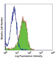
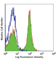
C57BL/6 mouse splenocytes were stained with CD18 (clone M18/2) Alexa Fluor® 594 (filled histogram) or rat IgG2a, κ Alexa Fluor® 594 isotype control (open histogram). The data was acquired by BD LSRFortessa™ cell analyzer equipped with Yellow-Green Laser (561 nm). -

Paraformaldehyde-fixed (4%), 500 µm-thick mouse spleen tissue section was processed according to the Ce3DTM Tissue Clearing Kit protocol (cat. no. 427701). The section was costained with anti-mouse/human CD45R/B220 Antibody (clone RA3-6B2) Alexa Fluor® 488 at 5 µg/mL (green), anti-mouse CD18 Antibody (clone M18/2) Alexa Fluor® 594 at 5 µg/mL (yellow), and anti-mouse CD4 Antibody (clone RM4-5) Alexa Fluor® 647 at 5 µg/mL (magenta). The section was then optically cleared and mounted in a sample chamber. The image was captured with a 10X objective using Zeiss 780 confocal microscope and processed by Imaris image analysis software.
Watch the video.
| Cat # | Size | Price | Quantity Check Availability | Save | ||
|---|---|---|---|---|---|---|
| 101416 | 100 µg | 203€ | ||||
CD18 is a 95 kD protein, also known as integrin β2 subunit. It is expressed on all leukocytes. CD18, in association with integrin α chain CD11a, CD11b, and CD11c forms LFA-1, Mac-1, and αXβ2, respectively, and plays an important role in leukocytes adhesion. The CD18 integrin complexes bind ICAM-1 (CD54), ICAM-2 (CD102), ICAM-3 (CD50), iC3b, and fibrinogen. The M18/2 antibody is able to block tumor cell metastasis and enhance cell adhesion.
Product DetailsProduct Details
- Verified Reactivity
- Mouse
- Antibody Type
- Monoclonal
- Host Species
- Rat
- Immunogen
- Mouse CTL glycoprotein
- Formulation
- Phosphate-buffered solution, pH 7.2, containing 0.09% sodium azide.
- Preparation
- The antibody was purified by affinity chromatography and conjugated with Alexa Fluor® 594 under optimal conditions.
- Concentration
- 0.5 mg/mL
- Storage & Handling
- The antibody solution should be stored undiluted between 2°C and 8°C, and protected from prolonged exposure to light. Do not freeze.
- Application
-
IHC-F - Quality tested
FC, 3D IHC- Verified - Recommended Usage
-
Each lot of this antibody is quality control tested by immunohistochemical staining on frozen tissue sections. For immunohistochemistry, a concentration range of 2.0 - 5.0 μg/mL is suggested. For flow cytometric staining, the suggested use of this reagent is ≤ 1.0 µg per million cells in 100 µL volume. For 3D immunohistochemistry on formalin-fixed tissues, a concentration of 5.0 µg/mL is suggested. It is recommended that the reagent be titrated for optimal performance for each application.
* Alexa Fluor® 594 has an excitation maximum of 590 nm, and a maximum emission of 617 nm.
Alexa Fluor® and Pacific Blue™ are trademarks of Life Technologies Corporation.
View full statement regarding label licenses - Excitation Laser
-
Green Laser (532 nm)/Yellow-Green Laser (561 nm)
- Application Notes
-
Additional reported applications (for the relevant formats) include: immunoprecipitation1,2, Western blotting1, in vitro and in vivo blocking of tumor cells metastasis or stimulation of cell-adhesion2-6, and immunohistochemical staining7 of acetone-fixed frozen sections and formalin-fixed paraffin-embedded sections. The Ultra-LEAF™ purified antibody (Endotoxin <0.01 EU/µg, Azide-Free, 0.2 µm filtered) is recommended for functional assays (Cat. No. 101417-101422).
-
Application References
(PubMed link indicates BioLegend citation) -
- Sanchez-Madrid, et al. 1983. J. Exp. Med. 158:586. (IP, WB)
- Zahalka MA, et al. 1993. J. Immunol. 150:4466. (IP, Block, Stim)
- Zahalka MA, et al. 1994. Int. Immunol. 6:917. (Block, Stim)
- Ishida Y, et al. 1994. Cell. Immunol. 155:414. (Block, Stim)
- Isobe M, et al. 1992. Science 255:1125. (Block, Stim)
- Driessens MHE, et al. 1996. J. Leukoc. Biol. 60:758. (Block, Stim)
- Barlow SC, et al. 2004. Amer. J. Pathol. 165:1849. (IHC)
- Product Citations
-
- RRID
-
AB_2563302 (BioLegend Cat. No. 101416)
Antigen Details
- Structure
- Integrin family, associates with CD11a, CD11b, CD11c, 95 kD
- Distribution
-
All leukocytes
- Function
- Adhesion
- Ligand/Receptor
- ICAM-1 (CD54), ICAM-2 (CD102), ICAM-3 (CD50), iC3b, fibrinogen
- Cell Type
- Leukocytes, Tregs
- Biology Area
- Cell Adhesion, Cell Biology, Immunology, Innate Immunity
- Molecular Family
- Adhesion Molecules, CD Molecules
- Antigen References
-
1. Barclay AN, et al. 1997. The Leukocyte Antigen FactsBook Academic Press.
2. Anderson DC, et al. 1987. Annu. Rev. Med. 38:175.
3. Wilson RW, et al. 1993. J. Immunol. 151:1571. - Gene ID
- 16414 View all products for this Gene ID
- UniProt
- View information about CD18 on UniProt.org
Related Pages & Pathways
Pathways


Related FAQs
Other Formats
View All CD18 Reagents Request Custom Conjugation| Description | Clone | Applications |
|---|---|---|
| FITC anti-mouse CD18 | M18/2 | FC |
| PE anti-mouse CD18 | M18/2 | FC |
| Purified anti-mouse CD18 | M18/2 | FC,IHC,IP,WB |
| Alexa Fluor® 647 anti-mouse CD18 | M18/2 | FC,3D IHC |
| Alexa Fluor® 594 anti-mouse CD18 | M18/2 | IHC-F,FC,3D IHC |
| Ultra-LEAF™ Purified anti-mouse CD18 | M18/2 | FC,IP,WB,Block,Stim,IHC |
Customers Also Purchased
Compare Data Across All Formats
This data display is provided for general comparisons between formats.
Your actual data may vary due to variations in samples, target cells, instruments and their settings, staining conditions, and other factors.
If you need assistance with selecting the best format contact our expert technical support team.
-
FITC anti-mouse CD18

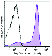
C57BL/6 mouse splenocytes stained with M18/2 FITC -
PE anti-mouse CD18

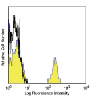
C57BL/6 splenocytes stained with M18/2 PE -
Purified anti-mouse CD18

C57BL/6 mouse splenocytes stained with purified M18/2, follo... -
Alexa Fluor® 647 anti-mouse CD18

C57BL/6 mouse splenocytes stained with M18/2 Alexa Fluor® 64... 
Paraformaldehyde-fixed (4%), 500 μm-thick mouse spleen tissu... -
Alexa Fluor® 594 anti-mouse CD18

C57BL/6 mouse frozen spleen section was fixed with 4% parafo... 
C57BL/6 mouse splenocytes were stained with CD18 (clone M18/... 
Paraformaldehyde-fixed (4%), 500 µm-thick mouse spleen tissu... -
Ultra-LEAF™ Purified anti-mouse CD18

C57BL/6 mouse splenocytes stained with Ultra-LEAF™ purified ...


Follow Us