 Login / Register


- Clone
- Poly19098 (See other available formats)
- Regulatory Status
- RUO
- Other Names
- Myosin-9, NMMHC-A, MYH9 variant protein, myosin heavy chain 9, non-muscle myosin heavy chain A, cellular myosin heavy chain, type A, non-muscle myosin heavy polypeptide 9
- Previously
-
Covance Catalog# PRB-440P
- Isotype
- Rabbit Polyclonal IgG
- Ave. Rating
- Submit a Review
- Product Citations
- publications

-

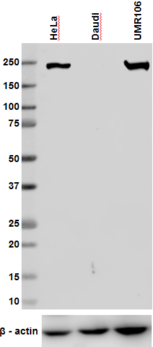
Western blot analysis of cell lysates from A431, Jurkat, THP-1 (Human), Raw264.7 (Mouse) and UMR106 (Rat) cells using Non-muscle Myosin Heavy Chain II-A (Myosin-9) Rabbit primary antibody (clone Poly19098, 1:100 dilution, 1 µg/ml) and HRP Donkey anti-Rabbit secondary antibody (Cat. No. 406401, 1:3000 dilution). Direct-Blot™ HRP anti-β-actin (Cat. No. 643807) was used as a loading control (1:8000 dilution). -

Immunofluorescence of HepG2 cells with (A) Rabbit Isotype control (Cat. No. 910801) or (B-D) Non-muscle Myosin Heavy Chain II-A (Myosin-9) primary antibody (clone19098). 2 µg/ml (1:250 dilution) Alexa Fluor® 594 (red) Donkey anti-Rabbit IgG (Cat. No. 406418) was used as secondary antibody. Nuclei were counterstained with DAPI (blue, Cat. No. 422801). The image was captured with a 60X objective using KEYENCE BZ-X700 fluorescence microscope. Exposure time (Seconds) for (A) is 1/30, and (B-D) is 1/40. Non-muscle Myosin Heavy Chain II-A antibody concentrations for (A, B) is 2 µg/ml (1:500 dilution), (C) is 1 µg/ml, (1:1000 dilution), (D) is 0.5 µg/ml. (1:500 dilution).
| Cat # | Size | Price | Quantity Check Availability | Save | ||
|---|---|---|---|---|---|---|
| 909802 | 25 µL | 72€ | ||||
| 909801 | 100 µL | 231€ | ||||
Non-muscle Myosin Heavy Chain II-A is a member of the myosin superfamily. There are three forms of myosin II, called myosin IIA, myosin IIB and myosin IIC. These proteins are involved in cell motility, maintenance of cell shape, and cytokinesis. Each type of myosin II protein consists of two heavy chains and four light chains. The heavy chains each have two parts: a myosin head-like domain and a tail region. The head region interacts with actin, a protein that is important for cell movement and shape. The long tail region interacts with other proteins, including the tail regions of other myosin proteins.
Product DetailsProduct Details
- Verified Reactivity
- Human, Mouse, Rat
- Reported Reactivity
- Cow
- Antibody Type
- Polyclonal
- Host Species
- Rabbit
- Immunogen
- This antibody was raised against the peptide GKADGAEAKPAE corresponding to the C-terminus of human nonmuscle myosin heavy chain isoform A (MHC-A, NMHC IIA, Myosin-9).
- Formulation
- Phosphate-buffered solution + 0.03% Thimerosal.
- Preparation
- The antibody was purified by affinity chromatography.
- Concentration
- 1 mg/ml
- Storage & Handling
- The antibody solution should be stored undiluted between 2°C and 8°C. Please note the storage condition for this antibody has been changed from -20°C to between 2°C and 8°C. You can also check your vial or your CoA to find the most accurate storage condition for this antibody.
- Application
-
WB - Quality tested
ICC - Verified - Recommended Usage
-
Each lot of this antibody is quality control tested by Western blotting. For Western blotting, the suggested use of this reagent is 1.0 - 2.0 µg per ml (1:500 - 1:1000 dilution). For immunocytochemistry, a concentration range of 0.5 - 1.0 μg/ml (1:500 - 1:1000 dilution) is recommended. The recommended usage for this polyclonal antibody may vary from lot to lot. It is recommended that the reagent be titrated for optimal performance for each application.
- Application Notes
-
This antibody is effective in immunoblotting (WB) and immunofluorescence (IF).
*Predicted MW = 200 kD
This product may contain other non-IgG subtypes. -
Application References
(PubMed link indicates BioLegend citation) -
- Hirota Y, et al.. 2010. Development 137: 3037. (IF) PubMed
- Ma X, et al. 2007. Mol Biol Cell. 18:2305. (IF)
- Golomb E, et al. 2004. J Biol Chem. 279:2800. (WB, IF)
- Song J, et al. 2001. Mol Cell Neurosci. 17:624.
- Kelley CA, et al. 1996. J Cell Biol. 134:675.
- Kelley CA, et al. 1995. J Biol Chem. 270:1395.
- Wang H, et al. 2008. J. Cell Biol. 182: 1171. (ICC, WB) PubMed
- Product Citations
-
- RRID
-
AB_2734686 (BioLegend Cat. No. 909802)
AB_2565100 (BioLegend Cat. No. 909801)
Antigen Details
- Biology Area
- Cell Biology, Cell Motility/Cytoskeleton/Structure, Neuroscience, Neuroscience Cell Markers
- Gene ID
- 4627 View all products for this Gene ID
- UniProt
- View information about Myosin-9 on UniProt.org

Other Formats
View All Myosin-9 Reagents Request Custom Conjugation| Description | Clone | Applications |
|---|---|---|
| Purified Non-muscle Myosin Heavy Chain II-A | Poly19098 | WB,ICC |
Customers Also Purchased
Compare Data Across All Formats
This data display is provided for general comparisons between formats.
Your actual data may vary due to variations in samples, target cells, instruments and their settings, staining conditions, and other factors.
If you need assistance with selecting the best format contact our expert technical support team.
-
Purified Non-muscle Myosin Heavy Chain II-A

Western blot analysis of cell lysates from A431, Jurkat, THP... 
Immunofluorescence of HepG2 cells with (A) Rabbit Isotype co...


Follow Us