 Login / Register


- Clone
- DO-1 (See other available formats)
- Regulatory Status
- RUO
- Other Names
- Tumor protein p53 (TP53)
- Isotype
- Mouse IgG2a, κ
- Ave. Rating
- Submit a Review
- Product Citations
- publications

-

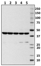
Whole cell extracts (15 µg total protein) from HEK293, A431, and THP-1 were resolved by 4-12% Bis-Tris gel electrophoresis, transferred to PVDF, and probed with 1.0 µg/mL (1:500 dilution) of Purified anti-p53 Antibody, clone DO-1, overnight at 4°C. Proteins were visualized by chemiluminescence detection using HRP goat anti-mouse IgG Antibody (Cat. No. 405306) at a 1:3000 dilution. Direct-Blot™ HRP anti-GAPDH Antibody (Cat. No. 607904) was used as a loading control at a 1:25000 dilution (lower). Lane M: Molecular Weight marker. Cell lysates were loaded in descending of predicted p53 expression; Predicted expression data was obtained from Human Protein Atlas. -

Immunofluorescence of Daudi cells with (A) Mouse IgG2b, κ Isotype control (Cat. No. 400302) or (B-D) TP-53 primary antibody. Alexa Fluor® 594 (Red) Goat anti-Mouse IgG (Cat. No. 405326) was used as secondary antibody. Nuclei were counterstained with DAPI (Blue, Cat. No. 422801). The image was captured with a 60X objective. Exposure time (Seconds) is 1/12. Concentrations for (A, B) is 5 µg/ml, (C) is 2 µg/ml, (D) is 1 µg/ml. -

Chromatin Immunoprecipitations (ChIP) were done with fixed and sonicated chromatin samples from 293T cells treated with tunicamycin (2 µg/mL, 8 hr). ChIP was performed with 5 µg of chromatin and either 1 µg of Go-ChIP-Grade™ purified anti-p53 antibody (Clone DO-1), or equal amount of Go-ChIP-Grade™ purified mouse IgG2a, κ isotype ctrl antibody (Clone MG2a-53, Cat. No. 401506). The enriched DNA was purified and quantified by real-time qPCR using SYBR Green and primers for the human CDKN1A promoter, and for a gene desert region on human chromosome 12. The amount of immunoprecipitated DNA in each sample is represented as a percentage of the total amount of input chromatin, which is equivalent to 100%.
| Cat # | Size | Price | Quantity Check Availability | Save | ||
|---|---|---|---|---|---|---|
| 645701 | 25 µg | 85€ | ||||
| 645702 | 100 µg | 181€ | ||||
p53 is a 53 kD protein that forms tetramers and functions as a tumor suppressor and transcriptional activator of genes that inhibit growth and/or invasion, cell cycle checkpoint after irradiation, DNA repair, apoptotic induction, signal transduction, and cell adhesion. This protein is localized to the nucleus when activated and can be upregulated by genotoxic or other cellular stresses. p53 is modified by phosphorylation, acetylation, ribosylation, ubiquitination, and sumoylation; ubiquination targets p53 for degradation via mdm2. This protein interacts with a variety of proteins including mdm2, mdmx, topoisomerase I, PML3, Bcl-XL, Bcl-2, Chk1, JNK, p38, HIPK2, CK2, DNA-PK, p300/CBP, PCAF, PARP1, and HDAC1-3. Mutant p53 associates with p63 and p73.
Product DetailsProduct Details
- Verified Reactivity
- Human
- Antibody Type
- Monoclonal
- Host Species
- Mouse
- Immunogen
- P53 N-terminal linear epitope aa 20-25
- Formulation
- This antibody is provided in phosphate-buffered solution containing 0.09% sodium azide.
- Preparation
- The antibody was purified by affinity chromatography.
- Concentration
- 0.5 mg/ml
- Storage & Handling
- The antibody solution should be stored undiluted between 2°C and 8°C.
- Application
-
WB - Quality tested
ICC, ChIP - Verified
IP - Reported in the literature, not verified in house - Recommended Usage
-
Each lot of this antibody is quality control tested by Western blotting. Western blotting, suggested working dilution(s): Use 0.5 - 1 µg per ml antibody for each mini-gel. For immunocytochemistry, a concentration range of 2.0 - 5.0 μg/ml (Dilution 1:100 - 1:250) is recommended. For ChIP applications, the recommended starting antibody to chromatin ratio is 1 µg of antibody and 5 µg of chromatin per IP. Optimal antibody to chromatin ratio should be determined by the end user. It is recommended that the reagent be titrated for optimal performance for each application.
-
Application References
(PubMed link indicates BioLegend citation) -
- Vojtesek B, et al. 1992. J Immuno Methods 151:237
- Stephen CW, et al. 1995. J Mol Biol 248:58
- Product Citations
-
- RRID
-
AB_1595606 (BioLegend Cat. No. 645701)
AB_2206462 (BioLegend Cat. No. 645702)
Antigen Details
- Structure
- Tetramer; 53 kD
- Distribution
-
Nuclear when activated
- Function
- Tumor suppressor, transcription factor regulates cell cycle checkpoint, apoptosis, DNA integrity
- Interaction
- mdm2, mdmx, TopoI, PML3, Bcl-XL, Bcl-2, Chk1, JNK, p38, HIPK2, CK2, DNA-PK, p300/CBP, PCAF, PARP1, HDAC1-3; mutant p53 associates with p63, p73
- Cell Type
- Mesenchymal Stem Cells
- Biology Area
- Cell Biology, DNA Repair/Replication, Stem Cells, Transcription Factors
- Antigen References
-
1. Vogelstein B, et al. 1992. Cell. 70:523.
2. Shieh S, et al. 1997. Cell. 91:325.
3. Mihara M, et al. 2003. Mol. Cell 11:577.
4. Saito S, et al. 2003. J. Biol. Chem. 278:37536. - Gene ID
- 7157 View all products for this Gene ID
- UniProt
- View information about p53 on UniProt.org

Related FAQs
Other Formats
View All p53 Reagents Request Custom Conjugation| Description | Clone | Applications |
|---|---|---|
| Purified anti-p53 | DO-1 | WB,ICC,IP,ChIP |
| Alexa Fluor® 594 anti-p53 | DO-1 | ICC |
Customers Also Purchased
Compare Data Across All Formats
This data display is provided for general comparisons between formats.
Your actual data may vary due to variations in samples, target cells, instruments and their settings, staining conditions, and other factors.
If you need assistance with selecting the best format contact our expert technical support team.


Follow Us