 Login / Register


- Clone
- Gal397 (See other available formats)
- Regulatory Status
- RUO
- Other Names
- Galectin-3, Mac-2, Gal-3, RL-29, galactose-specific lectin 3, CBP-35, L-34, εBP
- Isotype
- Mouse IgG1, κ
- Ave. Rating
- Submit a Review
- Product Citations
- publications

-

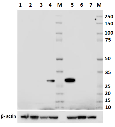
Total cell lysate from MCF7 cells (lane 1, 15 µg), Jurkat cells (lane 2, 15 µg), 3T3-L1 (lane 3, 15 µg) and Raw264.7 (lane 4, 15 µg) were resolved by electrophoresis (4-20% Tris-Glycine gel), transferred to nitrocellulose, and probed with purified anti-mouse/human Galectin-3 (Mac-2) antibody (clone Gal397). Proteins were visualized using an HRP Goat anti-mouse IgG Antibody and chemiluminescence detection. Direct-Blot™ HRP anti-β-actin antibody (clone 2F1-1) was used as a loading control. -

Total cell lysates (15 µg protein) from NIH3T3 (lane 1), HT29 (lane 2) and 293E (lane 3) cells were resolved by electrophoresis (4-20% Tris-Glycine gel), transferred to nitrocellulose, and probed with 1 µg/mL (1:500 dilution) of purified anti-mouse/human Galectin-3 (Mac-2) antibody (clone Gal397) (upper blot). Proteins were visualized by chemiluminescence detection using a 1:3000 diluted goat anti-mouse-IgG secondary antibody conjugated to HRP for anti-mouse/human Galectin-3 (Mac-2) antibody or 1:5000 diluted Direct-Blot HRP anti-β-Actin antibody, clone 2F1-1(lower blot). Lane M: Molecular weight ladder. -

HeLa cells were stained with purified anti-Galectin 3 (Gal397) antibody, followed by staining with DyLight™ 594 conjugated goat anti-mouse IgG (red) antibody. Actin filaments were labeled in green. Nuclei were stained with DAPI (blue).
| Cat # | Size | Price | Quantity Check Availability | Save | ||
|---|---|---|---|---|---|---|
| 126701 | 50 µg | 57€ | ||||
| 126702 | 500 µg | 207€ | ||||
Galectin-3 (galactose-specific lectin 3) is a glycoprotein, also known as Mac-2, Gal-3, RL-29, galactose-specific lectin 3, CBP-35, L-34, and εBP. It is a member of animal β-galactoside-binding protein family that is expressed in cell nucleus, cytoplasm, plasma membrane, or extracellular matrix by variety of tumor cells, monocytes/macrophages, epithelial cells, Kupffer cells, and dendritic cells. Galectin-3 contains carbohydrate recognition domains and binds to β-galactoside residues bearing glycoproteins, such as as IgE, IgA, galactose, casein kinase I, laminin, mucin, LAMPs, and CD66. Galectin-3 is an adhesion molecule and plays an important role in regulation of cell proliferation, differentiation and tumor cell metastasis. Galectin-3 structurely possesses NWGR motif that is conserved in BH-1 domain of BCL-2 family and functions as an anti-apoptotic molecule.
Product DetailsProduct Details
- Verified Reactivity
- Mouse, Human
- Reported Reactivity
- African Green, Baboon, Cynomolgus
- Antibody Type
- Monoclonal
- Host Species
- Mouse
- Immunogen
- Human Recombinant (partial), amino acids 151-251
- Formulation
- Phosphate-buffered solution, pH 7.2, containing 0.09% sodium azide.
- Preparation
- The antibody was purified by affinity chromatography.
- Concentration
- 0.5 mg/mL
- Storage & Handling
- The antibody solution should be stored undiluted between 2°C and 8°C.
- Application
-
WB - Quality tested
ICC - Verified - Recommended Usage
-
Each lot of this antibody is quality control tested by Western blotting. For Western blotting, the suggested use of this reagent is 1.0 µg per ml. For immunocytochemistry, a concentration of < 0.5 μg/ml is recommended. It is recommended that the reagent be titrated for optimal performance for each application.
- Application Notes
-
Based on immunogen information, clone Gal397 may recognize African Green Baboon and Cynomolgus. It is recommended to test the reagent for optimal performance for other species. Due to in-house testing, the purified format is not recommended for flow cytometry and intracellular flow cytometry.
- Product Citations
-
- RRID
-
AB_1134255 (BioLegend Cat. No. 126701)
AB_1134256 (BioLegend Cat. No. 126702)
Antigen Details
- Structure
-
Human Galetcin-3 has 250 amino acids with a predicted molecular weight of 26 kD.
Mouse Galetcin-3 has 264 amino acids with a predicted molecular weight of 27.5 kD. - Distribution
-
Localized in the nucleus, cytoplasm, cell surface, or extracellular matrix. Expressed by tumor cells, monocytes/macrophages, epithelial cells, Kupffer cells, dendritic cells
- Function
- Regulate cell proliferation and differentiation, adhesion, anti-apoptosis
- Ligand/Receptor
- Variety ligands, such as IgE, IgA, galactose, casein kinase I, laminin, mucin, LAMPs, CD66, etc
- Cell Type
- Dendritic cells, Epithelial cells, Macrophages, Monocytes
- Biology Area
- Cell Adhesion, Cell Biology, Cell Cycle/DNA Replication, Immunology, Innate Immunity
- Molecular Family
- Adhesion Molecules
- Antigen References
-
- Kim HRC, et al. 1999. Cancer Res. 59:4148
- Reljic R, et al. 2004. Immunol. Lett. 93:51.
- Takenaka Y et al. 2004. Mol. and Cell. Biol. 24:4395
- Joo GH, et al. 2001. J. Leukoc. Biol. 69:555
- Llinas L, et al. 2011. Immunol. Lett. 134:113-21
- Cabezón R, et al. 2011. Immunol. Lett. 134:167-73
- Gene ID
- 16854 View all products for this Gene ID 3958 View all products for this Gene ID
- UniProt
- View information about Mac-2 on UniProt.org

Related FAQs
Other Formats
View All Mac-2 Reagents Request Custom Conjugation| Description | Clone | Applications |
|---|---|---|
| PE anti-mouse/human Galectin-3 (Mac-2) | Gal397 | ICFC |
| Purified anti-mouse/human Galectin-3 (Mac-2) | Gal397 | WB,ICC |
Customers Also Purchased
Compare Data Across All Formats
This data display is provided for general comparisons between formats.
Your actual data may vary due to variations in samples, target cells, instruments and their settings, staining conditions, and other factors.
If you need assistance with selecting the best format contact our expert technical support team.
-
PE anti-mouse/human Galectin-3 (Mac-2)

Human peripheral blood monocytes intracellular stained with ... -
Purified anti-mouse/human Galectin-3 (Mac-2)

Total cell lysate from MCF7 cells (lane 1, 15 µg), Jurkat ce... 
Total cell lysates (15 µg protein) from NIH3T3 (lane 1), HT2... 
HeLa cells were stained with purified anti-Galectin 3 (Gal39...


Follow Us