 Login / Register


- Clone
- I3/2.3 (See other available formats)
- Regulatory Status
- RUO
- Other Names
- T200, Ly-5, LCA
- Isotype
- Rat IgG2b, λ
- Ave. Rating
- Submit a Review
- Product Citations
- publications

-

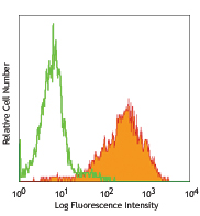
C57BL/6 splenocytes were stained with purified CD45 (clone I3/2.3) (filled histogram) or purified rat IgG2b isotype control (open histogram), followed by anti-rat IgG FITC.
| Cat # | Size | Price | Quantity Check Availability | Save | ||
|---|---|---|---|---|---|---|
| 147701 | 50 µg | 44€ | ||||
| 147702 | 500 µg | 175€ | ||||
CD45 is a 180-240 kD glycoprotein also known as the leukocyte common antigen (LCA), T200, or Ly-5. It is a member of the protein tyrosine phosphatase (PTP) family, expressed on all hematopoietic cells except mature erythrocytes and platelets. There are different isoforms of CD45 that arise from alternative splicing of exons 4, 5, and 6, which encode A, B, and C determinants, respectively. CD45 plays a key role in TCR and BCR signal transduction. These isoforms are very specific to the activation and maturation state of the cell as well as cell type. The primary ligands for CD45 are galectin-1, CD2, CD3, CD4, TCR, CD22, and Thy-1.
Product DetailsProduct Details
- Verified Reactivity
- Mouse
- Antibody Type
- Monoclonal
- Host Species
- Rat
- Immunogen
- Mouse lymphoma cell line
- Formulation
- Phosphate-buffered solution, pH 7.2, containing 0.09% sodium azide.
- Preparation
- The antibody was purified by affinity chromatography.
- Concentration
- 0.5 mg/ml
- Storage & Handling
- The antibody solution should be stored undiluted between 2°C and 8°C.
- Application
-
FC - Quality tested
IHC-P, IHC-F - Reported in the literature, not verified in house
SB - Community verified - Recommended Usage
-
Each lot of this antibody is quality control tested by immunofluorescent staining with flow cytometric analysis. For flow cytometric staining, the suggested use of this reagent is ≤ 0.125 µg per million cells in 100 µl volume. It is recommended that the reagent be titrated for optimal performance for each application.
- Application Notes
-
Additional reported applications (for the relevant formats) include: immunohistochemical staining of paraffin embedded sections1 and frozen tissue sections2.
- Additional Product Notes
-
This product has been verified for IHC-F (Immunohistochemistry - frozen tissue sections) on the NanoString GeoMx® Digital Spatial Profiler. The GeoMx® enables researchers to perform spatial analysis of protein and RNA targets in FFPE and fresh frozen human and mouse samples. For more information about our spatial biology products and the GeoMx® platform, please visit our spatial biology page.
-
Application References
(PubMed link indicates BioLegend citation) -
- Kliment C, et al. 2009. J. Mol. Cell Cardiol. 47:730. (IHC)
- Reynolds JM, et al. 2007. J. Immunol. 179:313. (IHC)
- Product Citations
-
- RRID
-
AB_2563415 (BioLegend Cat. No. 147701)
AB_2563416 (BioLegend Cat. No. 147702)
Antigen Details
- Structure
- Protein tyrosine phosphatase (PTP) family, 180-240 kD
- Distribution
-
All hematopoietic cells except mature erythrocytes and platelets
- Function
- Phosphatase, T and B cell activation
- Ligand/Receptor
- Galectin-1, CD2, CD3, CD4, TCR, CD22, Thy-1
- Cell Type
- B cells, Mesenchymal Stem Cells
- Biology Area
- Cell Biology, Immunology, Inhibitory Molecules, Neuroscience, Neuroscience Cell Markers, Stem Cells
- Molecular Family
- CD Molecules, TCRs
- Antigen References
-
1. Barclay A, et al. 1997. The Leukocyte Antigen FactsBook Academic Press.
2. Trowbridge IS and Thomas ML. 1994. Annu. Rev. Immunol. 12:85.
3. Kishihara K, et al. 1993. Cell 74:143.
4. Pulido R, et al. 1988. J. Immunol. 140:3851. - Gene ID
- 19264 View all products for this Gene ID
- UniProt
- View information about CD45 on UniProt.org


Related FAQs
- If an antibody clone has been previously successfully used in IBEX in one fluorescent format, will other antibody formats work as well?
-
It’s likely that other fluorophore conjugates to the same antibody clone will also be compatible with IBEX using the same sample fixation procedure. Ultimately a directly conjugated antibody’s utility in fluorescent imaging and IBEX may be specific to the sample and microscope being used in the experiment. Some antibody clone conjugates may perform better than others due to performance differences in non-specific binding, fluorophore brightness, and other biochemical properties unique to that conjugate.
- Will antibodies my lab is already using for fluorescent or chromogenic IHC work in IBEX?
-
Fundamentally, IBEX as a technique that works much in the same way as single antibody panels or single marker IF/IHC. If you’re already successfully using an antibody clone on a sample of interest, it is likely that clone will have utility in IBEX. It is expected some optimization and testing of different antibody fluorophore conjugates will be required to find a suitable format; however, legacy microscopy techniques like chromogenic IHC on fixed or frozen tissue is an excellent place to start looking for useful antibodies.
- Are other fluorophores compatible with IBEX?
-
Over 18 fluorescent formats have been screened for use in IBEX, however, it is likely that other fluorophores are able to be rapidly bleached in IBEX. If a fluorophore format is already suitable for your imaging platform it can be tested for compatibility in IBEX.
- The same antibody works in one tissue type but not another. What is happening?
-
Differences in tissue properties may impact both the ability of an antibody to bind its target specifically and impact the ability of a specific fluorophore conjugate to overcome the background fluorescent signal in a given tissue. Secondary stains, as well as testing multiple fluorescent conjugates of the same clone, may help to troubleshoot challenging targets or tissues. Using a reference control tissue may also give confidence in the specificity of your staining.
- How can I be sure the staining I’m seeing in my tissue is real?
-
In general, best practices for validating an antibody in traditional chromogenic or fluorescent IHC are applicable to IBEX. Please reference the Nature Methods review on antibody based multiplexed imaging for resources on validating antibodies for IBEX.
Other Formats
View All CD45 Reagents Request Custom Conjugation| Description | Clone | Applications |
|---|---|---|
| Purified anti-mouse CD45 | I3/2.3 | FC,IHC-P,IHC-F,SB |
| PerCP/Cyanine5.5 anti-mouse CD45 | I3/2.3 | FC |
| PE/Cyanine7 anti-mouse CD45 | I3/2.3 | FC |
| APC anti-mouse CD45 | I3/2.3 | FC |
| FITC anti-mouse CD45 | I3/2.3 | FC |
| PE anti-mouse CD45 | I3/2.3 | FC |
| APC/Fire™ 750 anti-mouse CD45 | I3/2.3 | FC |
| Alexa Fluor® 700 anti-mouse CD45 | I3/2.3 | FC |
| APC/Cyanine7 anti-mouse CD45 Antibody | I3/2.3 | FC |
Customers Also Purchased
Compare Data Across All Formats
This data display is provided for general comparisons between formats.
Your actual data may vary due to variations in samples, target cells, instruments and their settings, staining conditions, and other factors.
If you need assistance with selecting the best format contact our expert technical support team.
-
Purified anti-mouse CD45

C57BL/6 splenocytes were stained with purified CD45 (clone I... -
PerCP/Cyanine5.5 anti-mouse CD45

C57BL/6 splenocytes were stained with CD45 (clone I3/2.3) Pe... -
PE/Cyanine7 anti-mouse CD45

C57BL/6 splenocytes were stained with CD45 (clone I3/2.3) PE... -
APC anti-mouse CD45

C57BL/6 splenocytes were stained with CD45 (clone I3/2.3) AP... -
FITC anti-mouse CD45

C57BL/6 splenocytes were stained with CD45 (clone I3/2.3) FI... -
PE anti-mouse CD45

C57BL/6 splenocytes were stained with CD45 (clone I3/2.3) PE... -
APC/Fire™ 750 anti-mouse CD45

C57BL/6 mouse splenocytes were stained with CD45 (clone I3/2... -
Alexa Fluor® 700 anti-mouse CD45

C57BL/6 mouse splenocytes were stained with CD45 (clone I3/2... -
APC/Cyanine7 anti-mouse CD45 Antibody

C57BL/6 splenocytes were stained with anti-mouse CD45 (clone...


Follow Us