 Login / Register


- Clone
- Poly19053 (See other available formats)
- Regulatory Status
- RUO
- Other Names
- Keratin, type I cytoskeletal 14, CK-14, cytokeratin 14, keratin 14 (epidermolysis bullosa simplex, Dowling-Meara, Koebner)
- Isotype
- Rabbit Polyclonal
- Ave. Rating
- Submit a Review
- Product Citations
- publications

-

Western blot analysis of cell lysates from HaCaT (Positive control) and U87-MG (Low expression negative control) cells using Keratin 14 Rabbit primary antibody and HRP Donkey anti-Rabbit secondary antibody (#406401). Direct-Blot™ HRP anti-β-actin (#643807) was used as a loading control. -

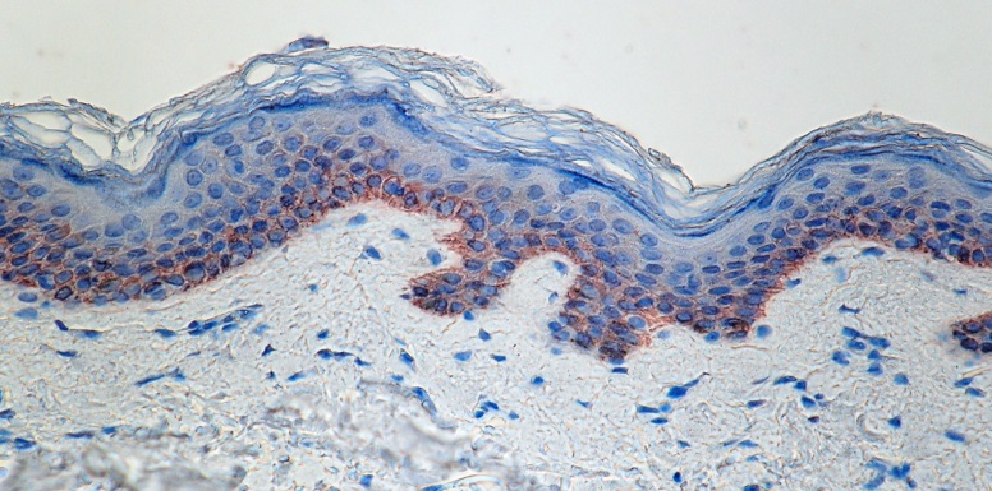
IHC staining of anti-Keratin 14 antibody (Poly19053) on formalin-fixed paraffin-embedded normal human skin. After antigen retrieval using Sodium Citrate, the tissue was incubated with the primary antibody at 1 µg/ml for 60 minutes at room temperature. BioLegend’s Ultra Streptavidin (USA) HRP Detection Kit was used for detection followed by hematoxylin counterstaining, according to the protocol provided. -

Western blot of anti-Keratin 14 antibody (Poly19053). Lane 1: Molecular weight marker; Lane 2: Human skin lysate. The blot was incubated with the primary antibody at 1 µg/ml, overnight at 4°C, followed by incubation with a fluorescent labeled goat anti-mouse secondary antibody. -

Whole mount mouse tail epidermis was stained with purified anti-Keratin 14 (green, clone Poly19053), purified anti-Keratin 15 (orange, clone Poly18339) and DAPI (blue). Image generously submitted to the 2017 Cell Life Imaging Competition by Kif Liakath. -

Immunofluorescence of HaCaT cells with (A) Rabbit Isotype control (Cat. No. 910801) or (B-D) Keratin 14 primary Antibody (Clone Poly 19053). 2 µg/ml (1:250 dilution) Alexa Fluor® 594 (red) Donkey anti-Rabbit IgG (Cat. No. 406418) was used as secondary antibody. Nuclei were counterstained with DAPI (blue). The image was captured with a 60X objective using KEYENCE BZ-X700 fluorescence microscope. Exposure time (Seconds) for (A) is 1/30, and (B-D) is 1/60. Anti-Keratin 14 primary antibody concentrations for (A, B) is 2 µg/ml (1:250 dilution), (C) is 1 µg/ml (1:1000 dilution), (D) is 0.5 µg/ml (1:1000 dilution).
| Cat # | Size | Price | Quantity Check Availability | Save | ||
|---|---|---|---|---|---|---|
| 905303 | 25 µg | 100€ | ||||
| 905304 | 100 µg | 280€ | ||||
Keratin 14 is a member of the keratin family, the most diverse group of intermediate filaments. This gene product, a type I keratin, is usually found as a heterotetramer with two keratin 5 molecules, a type II keratin. Together they form the cytoskeleton of epithelial cells. Mutations in the genes for these keratins are associated with epidermolysis bullosa simplex.
Product DetailsProduct Details
- Verified Reactivity
- Human, Mouse, Rat
- Reported Reactivity
- Dog, Non-Human Primate
- Antibody Type
- Polyclonal
- Host Species
- Rabbit
- Formulation
- Phosphate-buffered solution, pH 7.2, containing 0.09% sodium azide.
- Preparation
- The antibody was purified by affinity chromatography.
- Storage & Handling
- The antibody solution should be stored undiluted between 2°C and 8°C.
- Application
-
IHC-P - Quality tested
ICC, WB - Verified - Recommended Usage
-
Each lot of this antibody is quality control tested by formalin-fixed paraffin-embedded immunohistochemical staining. For immunohistochemistry, a concentration range of 0.1 - 10 μg/ml is suggested. For Western blotting, a concentration range of 0.5 - 2 ug/ml (1:250-1:1000) is suggested. For immunocytochemistry, a concentration range of 0.5 - 2.0 μg/ml (1:250 - 1:1000) is recommended. It is recommended that the reagent be titrated for optimal performance for each application.
- Application Notes
-
This product may contain other non-IgG subtypes. -
Application References
(PubMed link indicates BioLegend citation) - Product Citations
-
- RRID
-
AB_2734678 (BioLegend Cat. No. 905303)
AB_2616896 (BioLegend Cat. No. 905304)
Antigen Details
- Structure
- Keratin 14 is a 472 amino acid protein with a molecular mass of 52 kD.
- Distribution
-
Tissue distribution: skin.
Cellular distribution: extracellular, nucleus, cytoskeleton, and cytosol. - Function
- Nonhelical tail domain is involved in promoting KRT5-KRT14 filaments to self-organize into large bundles, enhances the mechanical properties involved in resilience of keratin intermediate filaments in vitro.
- Biology Area
- Cell Biology, Cell Motility/Cytoskeleton/Structure, Neuroscience, Neuroscience Cell Markers
- Molecular Family
- Intermediate Filaments
- Antigen References
-
1. Wend P, et al. 2013. EMBO J. 32(14):1977-89. PubMed
2. Welm AL, et al. 2005. Proc. Natl. Acad. Sci. USA 102(12):4324. (IHC) PubMed
3. Liang Y. 2011. Pathalog. Res. Int. 2011:93674. (IHC) PubMed - Gene ID
- 3861 View all products for this Gene ID
- UniProt
- View information about Keratin 14 on UniProt.org

Other Formats
View All Keratin 14 Reagents Request Custom Conjugation| Description | Clone | Applications |
|---|---|---|
| Keratin 14 Polyclonal Antibody, Purified | Poly19053 | IHC |
| Purified anti-Keratin 14 | Poly19053 | IHC-P,ICC,WB |
Customers Also Purchased
Compare Data Across All Formats
This data display is provided for general comparisons between formats.
Your actual data may vary due to variations in samples, target cells, instruments and their settings, staining conditions, and other factors.
If you need assistance with selecting the best format contact our expert technical support team.
-
Keratin 14 Polyclonal Antibody, Purified

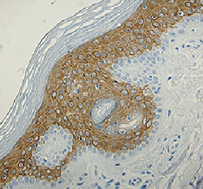
Strong cytoplasmic staining of the epidermis in normal human... 

-
Purified anti-Keratin 14

IHC staining of anti-Keratin 14 antibody (Poly19053) on form... 
Western blot of anti-Keratin 14 antibody (Poly19053). Lane 1... 
Western blot analysis of cell lysates from HaCaT (Positive c... 
Immunofluorescence of HaCaT cells with (A) Rabbit Isotype co... 
Whole mount mouse tail epidermis was stained with purified a...


Follow Us