 Login / Register


- Clone
- B1 (See other available formats)
- Regulatory Status
- RUO
- Other Names
- T cell receptor γ/δ, γ/δ TCR, TCR-γ/δ
- Isotype
- Mouse IgG1, κ
- Ave. Rating
- Submit a Review
- Product Citations
- publications

-

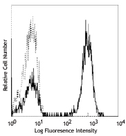
Human peripheral blood lymphocytes stained with purified B1, followed by anti-mouse IgG FITC
| Cat # | Size | Price | Quantity Check Availability | Save | ||
|---|---|---|---|---|---|---|
| 331202 | 100 µg | 92€ | ||||
T cell receptor (TCR) is a heterodimer consisting of an α and a β chain (TCR α/β) or a γ and a δ chain (TCR γ/δ). TCR γ/δ is involved in the recognition of certain bacterial, self-CD1 molecule, and tumor antigens bound to MHC class I. The γ/δ TCR associates with CD3 and is expressed on a subset of T cells found in the thymus, the intestinal epithelium, and the peripheral lymphoid tissues and peritoneum. Most γ/δ T cells are CD4-/CD8-, some are CD8+. T cells expressing the γ/δ TCR have been shown to play a role in oral tolerance, innate immune response for some tumor cells, and autoimmune disease. It has been reported that γ/δ T cells also play a principal role in antigen presentation.
Product DetailsProduct Details
- Verified Reactivity
- Human, Cynomolgus, Rhesus
- Reported Reactivity
- African Green, Baboon, Pigtailed Macaque
- Antibody Type
- Monoclonal
- Host Species
- Mouse
- Formulation
- Phosphate-buffered solution, pH 7.2, containing 0.09% sodium azide.
- Preparation
- The antibody was purified by affinity chromatography.
- Concentration
- 0.5 mg/ml
- Storage & Handling
- The antibody solution should be stored undiluted between 2°C and 8°C.
- Application
-
FC - Quality tested
IHC - Reported in the literature, not verified in house - Recommended Usage
-
Each lot of this antibody is quality control tested by immunofluorescent staining with flow cytometric analysis. For flow cytometric staining, the suggested use of this reagent is ≤2.0 µg per million cells in 100 µl volume. It is recommended that the reagent be titrated for optimal performance for each application.
- Application Notes
-
Clone B1 is also known as clone B1.1.
Additional reported applications (for the relevant formats) include: immunohistochemical staining of acetone-fixed frozen sections3 and paraffin-embedded sections5, in vitro blocking, and spatial biology (IBEX)8,9. The Ultra-LEAF™ purified antibody (Endotoxin < 0.01 EU/µg, Azide-Free, 0.2 µm filtered) is recommended for highly sensitive assays (Cat. Nos. 331235 and 331236). -
Application References
(PubMed link indicates BioLegend citation) -
- Rodriguez-Gago M, et al. 2001. Transplantation. 72:503.
- Lehmann FS, et al. 2002. Am. J. Physiol. Gastrointest. Liver. Physiol. 283:G481. (FC)
- Bordignon M, et al. 2008. Mol. Med. Rep. 1:485. (IHC)
- Conrad M, et al. 2007. Cytom. Part A 71A:925. (FC)
- Pollinger B, et al. 2011. J. Immunol. 186:2602. (IHC)
- Correia DV, et al. 2011. Blood. 118:992. (Block)
- Laurent AJ, et al. 2014. PLoS One. 9:103683. PubMed
- Radtke AJ, et al. 2020. Proc Natl Acad Sci USA. 117:33455-33465. (SB) PubMed
- Radtke AJ, et al. 2022. Nat Protoc. 17:378-401. (SB) PubMed
- Product Citations
-
- RRID
-
AB_1089222 (BioLegend Cat. No. 331202)
Antigen Details
- Structure
- Ig superfamily, associates with CD3 complex
- Distribution
-
T subset in thymus, intestinal epithelium, peripheral lymphoid tissues and peritoneum
- Function
- Antigen recognition
- Ligand/Receptor
- Some bacterial or tumor antigens bound MHC class I, CD1 molecule
- Cell Type
- Epithelial cells, T cells
- Biology Area
- Adaptive Immunity, Immunology
- Molecular Family
- TCRs
- Antigen References
-
1. Lanier LL, et al. 1987. J. Clin. Immunol. 7:429.
2. Spencer J, et al. 1989. Eur. J. Immunol. 19:1335.
3. Uyemura K, et al. 1991. J. Exp. Med. 174:683.
4. Spada FM, et al. 2000. J. Exp. Med. 191:907. - Gene ID
- 6964 View all products for this Gene ID 6965 View all products for this Gene ID
- UniProt
- View information about TCR gamma/delta on UniProt.org

Related FAQs
Other Formats
View All TCR γ/δ Reagents Request Custom Conjugation| Description | Clone | Applications |
|---|---|---|
| Purified anti-human TCR γ/δ | B1 | FC,IHC |
| Biotin anti-human TCR γ/δ | B1 | FC,IHC |
| FITC anti-human TCR γ/δ | B1 | FC |
| PE anti-human TCR γ/δ | B1 | FC,SB |
| APC anti-human TCR γ/δ | B1 | FC |
| Alexa Fluor® 647 anti-human TCR γ/δ | B1 | FC |
| Brilliant Violet 421™ anti-human TCR γ/δ | B1 | FC |
| Brilliant Violet 510™ anti-human TCR γ/δ | B1 | FC |
| PE/Cyanine7 anti-human TCR γ/δ | B1 | FC |
| PerCP/Cyanine5.5 anti-human TCR γ/δ | B1 | FC |
| PE/Dazzle™ 594 anti-human TCR γ/δ | B1 | FC |
| APC/Fire™ 750 anti-human TCR γ/δ | B1 | FC |
| TotalSeq™-A0139 anti-human TCR γ/δ | B1 | PG |
| TotalSeq™-C0139 anti-human TCR γ/δ | B1 | PG |
| TotalSeq™-B0139 anti-human TCR γ/δ | B1 | PG |
| Ultra-LEAF™ Purified anti-human TCR γ/δ | B1 | FC,FA,IHC,Block |
| PE/Fire™ 700 anti-human TCR γ/δ Antibody | B1 | FC |
| Alexa Fluor® 660 anti-human TCR γ/δ Antibody | B1 | FC |
| TotalSeq™-D0139 anti-human TCR γ/δ | B1 | PG |
| PE/Fire™ 810 anti-human TCR γ/δ | B1 | FC |
| Brilliant Violet 650™ anti-human TCR γ/δ | B1 | FC |
| Brilliant Violet 785™ anti-human TCR γ/δ | B1 | FC |
Customers Also Purchased
Compare Data Across All Formats
This data display is provided for general comparisons between formats.
Your actual data may vary due to variations in samples, target cells, instruments and their settings, staining conditions, and other factors.
If you need assistance with selecting the best format contact our expert technical support team.
-
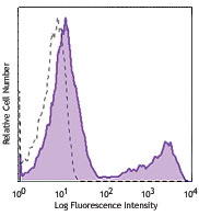
Purified anti-human TCR γ/δ

Human peripheral blood lymphocytes stained with purified B1,... -
Biotin anti-human TCR γ/δ

Human peripheral blood lymphocytes stained with CD3 APC and ... -
FITC anti-human TCR γ/δ


-
PE anti-human TCR γ/δ

Human peripheral blood lymphocytes stained with CD3 (UCHT1) ... 
Confocal image of C57BL/6 mouse thymus sample acquired using... -
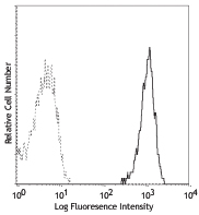
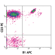
APC anti-human TCR γ/δ

Human peripheral blood lymphocytes stained with CD3 PE and B... -
Alexa Fluor® 647 anti-human TCR γ/δ

Human peripheral blood lymphocytes stained with CD3 PE and B... -
Brilliant Violet 421™ anti-human TCR γ/δ

Human peripheral blood lymphocytes were stained with CD3 APC... 
-
Brilliant Violet 510™ anti-human TCR γ/δ

Human peripheral blood lymphocytes were stained with CD3 APC... 
-
PE/Cyanine7 anti-human TCR γ/δ

Human peripheral blood lymphocytes were stained with CD3 FIT... -
PerCP/Cyanine5.5 anti-human TCR γ/δ

Human peripheral blood lymphocytes were stained with CD3 APC... -
PE/Dazzle™ 594 anti-human TCR γ/δ

Human peripheral blood lymphocytes were stained with CD3 APC... 
-
APC/Fire™ 750 anti-human TCR γ/δ

Human peripheral blood lymphocytes were stained with CD3 PE ... 
-
TotalSeq™-A0139 anti-human TCR γ/δ
-
TotalSeq™-C0139 anti-human TCR γ/δ
-
TotalSeq™-B0139 anti-human TCR γ/δ
-
Ultra-LEAF™ Purified anti-human TCR γ/δ
Human peripheral blood lymphocytes stained with purified B1,... -
PE/Fire™ 700 anti-human TCR γ/δ Antibody

Human peripheral blood lymphocytes were stained with anti-hu... -
Alexa Fluor® 660 anti-human TCR γ/δ Antibody

Human peripheral blood lymphocytes were stained with CD3 FIT... -
TotalSeq™-D0139 anti-human TCR γ/δ
-
PE/Fire™ 810 anti-human TCR γ/δ

Human peripheral blood lymphocytes were stained with anti-hu... -
Brilliant Violet 650™ anti-human TCR γ/δ

Human peripheral blood lymphocytes were stained with anti-hu... -
Brilliant Violet 785™ anti-human TCR γ/δ

Human peripheral blood lymphocytes were stained with anti-hu...


Follow Us