 Login / Register


- Clone
- C20Mab-60 (See other available formats)
- Regulatory Status
- RUO
- Other Names
- B1, Bp35
- Isotype
- Mouse IgG2a, κ
- Ave. Rating
- Submit a Review
- Product Citations
- publications

-

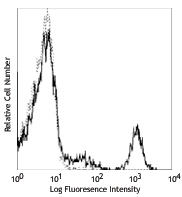
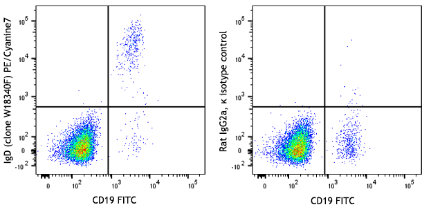
Human peripheral blood lymphocytes were surface stained with anti-human CD19 APC and purified anti-human CD20 (clone C20Mab-60) (left), or mouse IgG2a, κ isotype control (right) followed by PE goat anti-mouse IgG. -

Human paraffin-embedded spleen tissue slices were prepared with a standard protocol of deparaffination and rehydration. Antigen retrieval was done with 1X Tris-EDTA buffer (pH 9.0) at 95°C for 40 minutes. Tissue was washed with PBS/ 0.05% Tween-20 twice for five minutes, blocked with 5% FBS and 0.2% gelatin for 30 minutes. Then, the tissue was stained with 10 µg/mL of purified anti-human CD20 (clone C20Mab-60) and 5 µg/mL of purified anti-human CD4 (clone A17070D) at 4°C overnight. On the next day, the tissue was washed twice with PBS and stained with Alexa Fluor® 647 anti-mouse IgG2a (red) and Alexa Fluor® 594 anti-mouse IgG2b (green) for two hours at room temperature. The nuclei were counterstained with DAPI (blue). The image was captured with a 10X objective. -

Human paraffin-embedded tonsil tissue slices were prepared with a standard protocol of deparaffination and rehydration. Antigen retrieval was done with 1X Tris-EDTA buffer (pH 9.0) at 95°C for 40 minutes. Tissue was washed with PBS/ 0.05% Tween-20 twice for five minutes, blocked with 5% FBS and 0.2% gelatin for 30 minutes. Then, the tissue was stained with 10 µg/mL of purified anti-human CD20 (clone C20Mab-60) and 5 µg/mL of purified anti-human CD4 (clone A17070D) at 4°C overnight. On the next day, the tissue was washed twice with PBS and stained with Alexa Fluor® 647 anti-mouse IgG2a (red) and Alexa Fluor® 594 anti-mouse IgG2b (green) for two hours at room temperature. The nuclei were counterstained with DAPI (blue). The image was captured with a 10X objective. -
Human paraffin-embedded B cell lymphoma tissue slices were prepared with a standard protocol of deparaffination and rehydration. Antigen retrieval was done with 1X Tris-EDTA buffer (pH 9.0) at 95°C for 40 minutes. Tissue was washed with PBS/ 0.05% Tween-20 twice for five minutes, blocked with 5% FBS and 0.2% gelatin for 30 minutes. Then, the tissue was stained with 10 µg/mL of purified anti-human CD20 (clone C20Mab-60) at 4°C overnight. On the next day, the tissue was washed twice with PBS and stained with Alexa Fluor® 594 anti-mouse IgG (red) for two hours at room temperature. The nuclei were counterstained with DAPI (blue). The image was captured with a 10X objective.

| Cat # | Size | Price | Quantity Check Availability | Save | ||
|---|---|---|---|---|---|---|
| 382802 | 100 µg | 62€ | ||||
CD20 is a 33-37 kD, four transmembrane spanning protein, also known as B1 and Bp35. CD20 is expressed on pre-B-cells, resting and activated B cells (not plasma cells), some follicular dendritic cells, and at low levels on a T cell subset. CD20 is heavily phosphorylated on activated B cells and malignant B cells. Homo-oligomeric complexes of CD20 are thought to form Ca2+ conductive ion channels in the plasma membrane of B cells. The CD20 molecule is involved in B-cell activation and is associated with various Src family kinases (Lyn, Lck, Fyn). It exists in a complex with MHC class I and II, CD53, CD81, and CD82.
Product DetailsProduct Details
- Verified Reactivity
- Human
- Antibody Type
- Monoclonal
- Host Species
- Mouse
- Immunogen
- LN229/CD20 cells
- Formulation
- Phosphate-buffered solution, pH 7.2, containing 0.09% sodium azide
- Preparation
- The antibody was purified by affinity chromatography.
- Concentration
- 0.5 mg/mL
- Storage & Handling
- The antibody solution should be stored undiluted between 2°C and 8°C.
- Application
-
FC - Quality tested
IHC-P - Verified
WB - Reported in the literature, not verified in house - Recommended Usage
-
Each lot of this antibody is quality control tested by immunofluorescent staining with flow cytometric analysis. For flow cytometric staining, the suggested use of this reagent is ≤ 1.0 µg per million cells in 100 µL volume. For immunohistochemistry on formalin-fixed paraffin-embedded tissue sections, a concentration range of 5.0 - 10.0 µg/mL is suggested. It is recommended that the reagent be titrated for optimal performance for each application.
- RRID
-
AB_2924581 (BioLegend Cat. No. 382802)
Antigen Details
- Structure
- Four transmembrane protein (TM4SF), heavily phosphorylated after activation, 33-37 kD
- Distribution
-
B cell, T cell subsets
- Function
- B cell activation
- Ligand/Receptor
- Src family tyrosine kinases, MHC class I, II, CD53, CD81, CD82
- Cell Type
- B cells, T cells
- Biology Area
- Costimulatory Molecules, Immunology
- Molecular Family
- CD Molecules
- Antigen References
-
- Furusawa Y, et al. 2020. Monoclon Antib Immunodiagn Immunother. 39:112-116.
- Gene ID
- 931 View all products for this Gene ID
- UniProt
- View information about CD20 on UniProt.org


Related FAQs
Other Formats
View All CD20 Reagents Request Custom Conjugation| Description | Clone | Applications |
|---|---|---|
| Purified anti-human CD20 | C20Mab-60 | FC,IHC-P,WB |
| Alexa Fluor® 647 anti-human CD20 | C20Mab-60 | FC,IHC-P |
| Alexa Fluor® 594 anti-human CD20 | C20Mab-60 | IHC-P,FC |
Customers Also Purchased
Compare Data Across All Formats
This data display is provided for general comparisons between formats.
Your actual data may vary due to variations in samples, target cells, instruments and their settings, staining conditions, and other factors.
If you need assistance with selecting the best format contact our expert technical support team.
-
Purified anti-human CD20

Human peripheral blood lymphocytes were surface stained with... 
Human paraffin-embedded spleen tissue slices were prepared w... 
Human paraffin-embedded tonsil tissue slices were prepared w... Human paraffin-embedded B cell lymphoma tissue slices were p... -
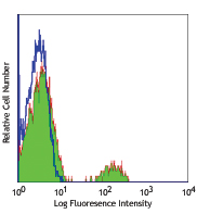
Alexa Fluor® 647 anti-human CD20

Human peripheral blood lymphocytes were stained with anti-hu... 
IHC staining of anti-human CD20 (clone C20Mab-60) Alexa Fluo... -
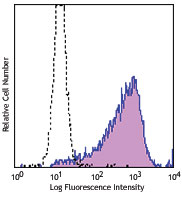
Alexa Fluor® 594 anti-human CD20

IHC staining of Alexa Fluor® 594 anti-human CD20 (clone ... 
Human peripheral blood lymphocytes were stained with anti-hu...


Follow Us