 Login / Register


- Clone
- STA (See other available formats)
- Regulatory Status
- RUO
- Workshop
- V A013
- Other Names
- VCAM-1, INCAM-110
- Isotype
- Mouse IgG1, κ
- Ave. Rating
- Submit a Review
- Product Citations
- publications

-

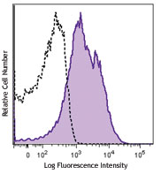
TNF-α stimulated HUVEC cells stained with purified STA, followed by anti-mouse IgGs FITC -

Human paraffin-embedded breast cancer tissue slices were prepared with a standard protocol of deparaffination and rehydration. Antigen retrieval was done with Tris-Buffered Saline 1X (1.0M, pH7.4) at 95°C for 40 minutes. Tissue was washed with PBS/ 0.05% Tween20 twice for five minutes and blocked with 5% FBS and 0.2% gelatin for 30 minutes. Then, the tissue was stained with 10 µg/ml of purified anti-human CD106 (clone STA) antibody overnight at 4°C. On the next day, the tissue was incubated with Alexa Fluor®594 Goat anti-mouse IgG (clone Poly4053) antibody (red) for an hour. Nuclei were counterstained with DAPI (blue). The image was captured with a 10X objective.
| Cat # | Size | Price | Quantity Check Availability | Save | ||
|---|---|---|---|---|---|---|
| 305802 | 100 µg | 81€ | ||||
CD106 is a 110 kD single chain type I glycoprotein also known as VCAM-1 and INCAM-110. It is expressed predominantly on activated vascular endothelium but has also been identified on follicular and interfollicular dendritic cells, some macrophages, bone marrow stromal cells, and non-vascular cell populations within joints, kidney, muscle, heart, placenta, and brain. Expression on endothelial cells as well as many other cells is induced by inflammatory stimuli and cytokines. Activated endothelial cells can release soluble forms of CD106 which can be detected in the blood. CD106 binds the integrins CD49d/CD29 (VLA-4) and α4β7 that contribute to leukocyte adhesion, transmigration, and co-stimulation of T cell proliferation.
Product DetailsProduct Details
- Verified Reactivity
- Human
- Antibody Type
- Monoclonal
- Host Species
- Mouse
- Formulation
- Phosphate-buffered solution, pH 7.2, containing 0.09% sodium azide.
- Preparation
- The antibody was purified by affinity chromatography.
- Concentration
- 0.5 mg/mL
- Storage & Handling
- The antibody solution should be stored undiluted between 2°C and 8°C.
- Application
-
FC - Quality tested
IHC-P - Verified
ICC, IP, ELISA - Reported in the literature, not verified in house - Recommended Usage
-
Each lot of this antibody is quality control tested by immunofluorescent staining with flow cytometric analysis. For flow cytometric staining, the suggested use of this reagent is ≤ 2.0 µg per million cells in 100 µl volume. For immunohistochemical staining on formalin-fixed paraffin-embedded tissue sections, the suggested use of this reagent is 5.0 - 10 µg per ml. It is recommended that the reagent be titrated for optimal performance for each application.
- Application Notes
-
Additional reported applications (for the relevant formats) include: immunofluorescence3, immunohistochemical staining of acetone-fixed frozen tissue sections, immunoprecipitation2, ELISA2 capture for sCD106, and spatial biology (IBEX)5,6.
-
Application References
(PubMed link indicates BioLegend citation) -
- Schlossman S, et al. Eds. 1995. Leucocyte Typing V. Oxford University Press. New York.
- Leca G, et al. 1995. J. Immunol. 154:1069. (ELISA IP)
- Yen YT, et al. 2006. J. Virol. 80:2648. (IF) PubMed
- Dmitrieva NI, et al. 2015. PloS One.10:128870. PubMed
- Radtke AJ, et al. 2020. Proc Natl Acad Sci USA. 117:33455-33465. (SB) PubMed
- Radtke AJ, et al. 2022. Nat Protoc. 17:378-401. (SB) PubMed
- Product Citations
-
- RRID
-
AB_314558 (BioLegend Cat. No. 305802)
Antigen Details
- Structure
- Ig superfamily, type I glycoprotein, 110 kD
- Distribution
-
Activated endothelial cells, endothelial progenitors, follicular dendritic cells
- Function
- Leukocyte adhesion, transmigration, costimulation
- Ligand/Receptor
- VLA-4 (CD49d/CD29)
- Cell Type
- Dendritic cells, Endothelial cells, Mesenchymal Stem Cells
- Biology Area
- Cell Adhesion, Cell Biology, Immunology, Neuroinflammation, Neuroscience, Stem Cells
- Molecular Family
- Adhesion Molecules, CD Molecules
- Antigen References
-
1. Carlos T, et al. 1994. Blood 84:2068.
2. Jones E, et al. 1995. Nature 373:539. - Gene ID
- 7412 View all products for this Gene ID
- UniProt
- View information about CD106 on UniProt.org


Related FAQs
Other Formats
View All CD106 Reagents Request Custom Conjugation| Description | Clone | Applications |
|---|---|---|
| APC anti-human CD106 | STA | FC |
| Biotin anti-human CD106 | STA | FC |
| PE anti-human CD106 | STA | FC,SB |
| PE/Cyanine5 anti-human CD106 | STA | FC |
| Purified anti-human CD106 | STA | FC,IHC-P,ICC,IP,ELISA |
| TotalSeq™-A0245 anti-human CD106 | STA | PG |
| PE/Cyanine7 anti-human CD106 | STA | FC |
| Brilliant Violet 421™ anti-human CD106 | STA | FC |
| TotalSeq™-C0245 anti-human CD106 | STA | PG |
| TotalSeq™-B0245 anti-human CD106 | STA | PG |
| KIRAVIA Blue 520™ anti-human CD106 | STA | FC |
Customers Also Purchased
Compare Data Across All Formats
This data display is provided for general comparisons between formats.
Your actual data may vary due to variations in samples, target cells, instruments and their settings, staining conditions, and other factors.
If you need assistance with selecting the best format contact our expert technical support team.
-
APC anti-human CD106

TNF-α stimulated HUVEC cells stained with STA APC -
Biotin anti-human CD106

TNF-α stimulated HUVEC cells stained with biotinylated STA,... -
PE anti-human CD106

TNF-a stimulated HUVEC cells stained with STA PE 
Confocal image of human lymph node sample acquired using the... 
Confocal image of human jejunum sample acquired using the IB... -
PE/Cyanine5 anti-human CD106

TNF-α stimulated HUVEC cells stained with STA PE/Cyani... -
Purified anti-human CD106

TNF-α stimulated HUVEC cells stained with purified STA, foll... 
Human paraffin-embedded breast cancer tissue slices were pre... -
TotalSeq™-A0245 anti-human CD106
-
PE/Cyanine7 anti-human CD106

TNF-a stimulated HUVEC cells stained with CD106 (clone STA) ... -
Brilliant Violet 421™ anti-human CD106

TNF-a stimulated HUVEC cells stained with CD106 (clone STA) ... -
TotalSeq™-C0245 anti-human CD106
-
TotalSeq™-B0245 anti-human CD106
-
KIRAVIA Blue 520™ anti-human CD106

TNF-α stimulated HUVEC cells stained with anti-human CD106 (...


Follow Us