 Login / Register


- Clone
- HTA28 (See other available formats)
- Regulatory Status
- RUO
- Other Names
- Histone-H3
- Isotype
- Rat IgG2a, κ
- Ave. Rating
- Submit a Review
- Product Citations
- publications

-

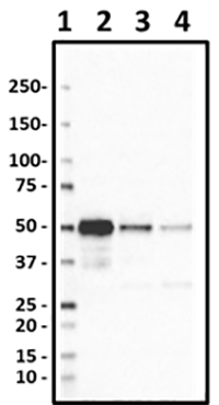
Whole cell extracts (15 µg protein) from untreated (-) or nocodazole-treated (+, 300 ng/mL overnight) HeLa cells were resolved on a 4-12% Bis-Tris gel, transferred to a nitrocellulose membrane and probed with 1.0 µg/mL (1:500 dilution) of Purified anti-Histone H3 Phospho (Ser28) Antibody, clone HTA28, overnight at 4°C. Proteins were visualized by chemiluminescence detection using HRP goat anti-mouse IgG Antibody (Cat. No. 405306) at a 1:3000 dilution. Direct-Blot™ anti-β-Actin was used as a loading control at a 1:25000 dilution. Lane M: Molecular Weight marker. -

HeLa cells were fixed with 4% paraformaldehyde (PFA) for 15 minutes, permeabilized with 0.5% Triton X-100 for 3 minutes, and blocked with 5% FBS for 60 minutes. Then the cells were intracellularly stained with 1:2500 diluted (0.2 µg/ml) Histone H3-Phosphorylated (Ser28) Antibody (clone HTA28, lower) overnight at 4 degrees followed by Alexa Fluor® 594 (red) conjugated goat anti-rat IgG (Cat. No. 405422) incubation for one hour at room temperature. Nuclei were counterstained with DAPI (blue). The image was captured with a 60X objective. The images displayed HeLa cells during cell cycle progression. -

Untreated HeLa (Panel A) and overnight nocodazoled-treated HeLa (Panel B) were stained with purified rat monoclonal antibody against phospho-H3 (Ser28) (clone HTA28), followed by Alexa Fluor® 488 anti-alpha-tubulin, DyLight™ 594 goat anti-rat-IgG and DAPI.
| Cat # | Size | Price | Quantity Check Availability | Save | ||
|---|---|---|---|---|---|---|
| 641001 | 25 µg | 141€ | ||||
| 641002 | 100 µg | 329€ | ||||
H3 is a core component of the nucleosome that serves to wrap and compact DNA into chromatin. Histones therefore, limit the accessibility of DNA, providing mechanisms for transcription regulation, DNA repair and replication and chromosomal stability. During mitosis, H3 is phosphorylated at serine 28.This phosphorylation coincides with chromosome condensation initiated at prophase and disappears at late anaphase. H3 has been demonstrated to be phosphorylated by the action of MLTK-α (mixed linage kinase-like mitogen activated protein triple kinase α) in response to ultraviolet B light and epidermal growth factor, as well as Aurora-B during mitosis.
Product DetailsProduct Details
- Verified Reactivity
- Human
- Antibody Type
- Monoclonal
- Host Species
- Rat
- Immunogen
- Synthetic peptide conjugated to KLH, corresponding to amino acids 23-35 of human histone H3.
- Formulation
- This antibody is provided in phosphate-buffered solution, pH 7.2, containing 0.09% sodium azide. Final antibody concentration is 0.5 mg/ml.
- Preparation
- The antibody was purified by affinity chromatography.
- Concentration
- 0.5 mg/ml
- Storage & Handling
- Upon receipt, store between 2°C and 8°C.
- Application
-
WB - Quality tested
CyTOF®, ICC - Verified
IP, ICFC - Reported in the literature, not verified in house - Recommended Usage
-
Each lot of this antibody is quality control tested by Western blotting. For Western blotting, the suggested use of this reagent is 0.5 - 1.0 µg per ml. It is recommended that the reagent be titrated for optimal performance for each application.
- Application Notes
-
This clone is not recommended for ChIP (Chromatin Immunoprecipitation) assays (as determined by in-house testing).
-
Application References
(PubMed link indicates BioLegend citation) -
- Hirata A, et al. 2004. J. Histochem. Cytochem. 52:1503.
- Goto H, et al. 1999. J. Biol. Chem. 274:25543.
- Ozawa K. 2008. Cytometry A 73:517.
- Goode NJ, et al. 2014. PLoS Genet. 10:1004323. PubMed
- Product Citations
-
- RRID
-
AB_1227660 (BioLegend Cat. No. 641001)
AB_1227659 (BioLegend Cat. No. 641002)
Antigen Details
- Structure
- H3 is part of the nucleosome, comprised of an octameric complex with H2A, H2B, and H4 proteins.
- Distribution
-
Nucleus
- Function
- H3 is a core component of the nucleosome that serves to wrap and compact DNA into chromatin. Histones therefore, limit the accessibility of DNA, providing mechanisms for transcription regulation, DNA repair and replication and chromosomal stability.
- Interaction
- Two molecules of H3 form a heterotetramer with two molecules of H4.
- Biology Area
- Cell Biology, DNA Repair/Replication, Transcription Factors
- Molecular Family
- Phospho-Proteins
- Antigen References
-
1. Choi HS, et al. 2005. J. Biol. Chem. 280:13545.
2. Goto H, et al. 2002. Genes Cells 7:11.
3. Garcia BA, et al. 2005. Biochemistry 44:13202. - Regulation
- H3 is regulated by acetylation, methylation, citrullination, phosphorylation, and ubiquitination.
- Gene ID
- 8290 View all products for this Gene ID
- UniProt
- View information about Histone on UniProt.org

Related FAQs
Other Formats
View All Histone Reagents Request Custom Conjugation| Description | Clone | Applications |
|---|---|---|
| Purified anti-Histone H3-Phosphorylated (Ser28) | HTA28 | WB,CyTOF®,ICC,IP,ICFC |
| Alexa Fluor® 488 anti-Histone H3-Phosphorylated (Ser28) | HTA28 | ICFC |
| Alexa Fluor® 647 anti-Histone H3-Phosphorylated (Ser28) | HTA28 | ICFC |
| Purified anti-Histone H3-Phosphorylated (Ser28) (Maxpar® Ready) | HTA28 | WB,CyTOF® |
| PE anti-Histone H3-Phosphorylated (Ser28) | HTA28 | ICFC |
| PE/Cyanine7 anti-Histone H3-Phosphorylated (Ser28) | HTA28 | ICFC |
| PerCP/Cyanine5.5 anti-Histone H3-Phosphorylated (Ser28) | HTA28 | ICFC |
| Alexa Fluor® 594 anti-Histone H3-Phosphorylated (Ser28) | HTA28 | ICC |
| Direct-Blot™ HRP anti-Histone H3 Phospho (Ser28) | HTA28 | WB |
Customers Also Purchased
Compare Data Across All Formats
This data display is provided for general comparisons between formats.
Your actual data may vary due to variations in samples, target cells, instruments and their settings, staining conditions, and other factors.
If you need assistance with selecting the best format contact our expert technical support team.
-
Purified anti-Histone H3-Phosphorylated (Ser28)

Whole cell extracts (15 µg protein) from untreated (-) or no... 
HeLa cells were fixed with 4% paraformaldehyde (PFA) for 15... 
Untreated HeLa (Panel A) and overnight nocodazoled-treated H... -
Alexa Fluor® 488 anti-Histone H3-Phosphorylated (Ser28)

Nocodazole-treated Hela cells intracellularly stained with H... -
Alexa Fluor® 647 anti-Histone H3-Phosphorylated (Ser28)

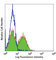
HELA cells (open histogram) and HELA cells treated with Noco... -
Purified anti-Histone H3-Phosphorylated (Ser28) (Maxpar® Ready)
-
PE anti-Histone H3-Phosphorylated (Ser28)

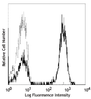
HeLa cells (open histogram) and HeLa cells treated with Noco... -
PE/Cyanine7 anti-Histone H3-Phosphorylated (Ser28)

HeLa cells (open histogram) and HeLa cells treated with Noco... -
PerCP/Cyanine5.5 anti-Histone H3-Phosphorylated (Ser28)

HeLa cells untreated (open histogram), and HeLa cells treate... -
Alexa Fluor® 594 anti-Histone H3-Phosphorylated (Ser28)

HeLa cells were were fixed with 4% paraformaldehyde (PFA) fo... -
Direct-Blot™ HRP anti-Histone H3 Phospho (Ser28)

Western blot analysis of 15µg cell lysates from HeLa with or...


Follow Us