 Login / Register


- Clone
- A18011A (See other available formats)
- Regulatory Status
- RUO
- Other Names
- Epidermal Growth Factor Receptor, Receptor Tyrosine-Protein Kinase ErbB-1, Erb-B2 Receptor Tyrosine Kinase 1, ERBB1, NISBD2, PIG61, MENA
- Isotype
- Mouse IgG1, κ
- Ave. Rating
- Submit a Review
- Product Citations
- publications

-

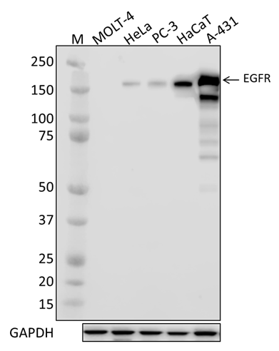
Whole cell extracts (15 µg protein) from MOLT-4 (negative control), HeLa, PC3, HaCaT, and A431 cells were resolved by 4-12% Bis-Tris gel electrophoresis, transferred to a PVDF membrane, and probed with 1.0 µg/mL (1:500 dilution) of Purified anti-EGFR antibody, clone A18011A, overnight at 4°C. Proteins were visualized by chemiluminescence detection using HRP goat anti-mouse IgG antibody (Cat. No. 405306) at a 1:3000 dilution. Direct-Blot™ HRP anti-GAPDH antibody (Cat. No. 607904) was used as a loading control at a 1:25000 dilution (lower). Lane M: Molecular Weight marker. Cell lysates were loaded in order of ascending EGFR expression; predicted expression data was obtained from Human Protein Atlas. -

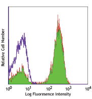
A431 cells were fixed with 4% paraformaldehyde for 10 minutes, permeabilized with Triton X-100 for 10 minutes, and blocked with 5% FBS for 60 minutes. Cells were then intracellularly stained with a 1:500 dilution (1.0 µg/mL) of either purified mouse IgG1, κ isotype control antibody (Cat. No. 401402, panel A) or purified anti-EGFR antibody (panel B) for two hours at room temperature, followed by incubation with Alexa Fluor™ 594 goat anti-mouse IgG (Cat. No. 405326) at 2.0 µg/mL. Nuclei were counter-stained with DAPI, and the image was captured with a 60X objective. -

Whole cell extracts (250 μg total protein) prepared from A431 cells were immunoprecipitated overnight with 2.5 µg of purified mouse IgG1, κ isotype control antibody (Cat. No. 401402) or purified anti-EGFR antibody, clone A18011A. The resulting IP fractions and whole cell extract input (6%) were resolved by 4-12% Bis-Tris gel electrophoresis, transferred to a PVDF membrane, and probed with a 0.1 µg/mL (1:5000 dilution) of A18011A. Lane M: Molecular Weight marker. -

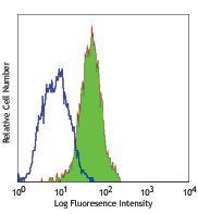
A-431 cells were stained intracellularly with purified EGFR (clone A18011A) (filled histogram) or mouse IgG1, κ isotype control (open histogram), followed by anti-mouse IgG PE.
| Cat # | Size | Price | Quantity Check Availability | Save | ||
|---|---|---|---|---|---|---|
| 617501 | 25 µg | 81€ | ||||
| 617502 | 100 µg | 203€ | ||||
Epidermal Growth Factor Receptor (EGFR) is a receptor tyrosine kinase that links extracellular mitogenic ligand binding to complex downstream signaling cascades. Initial ligand binding results in receptor oligomerization and autophosphorylation of multiple tyrosine residues within cytosolic domains of the the protein. These phosphorylation events stabilize the EGFR kinase activation loop and lead to the recruitment of adaptor proteins and other downstream effectors. EGFR stimulation leads to activation of RAS-RAF-MEK-ERK, PI3 kinase-AKT, PLCγ-PKC and STAT signaling cascades, and constitutive activation of the receptor promotes tumorigenesis in multiple cancers.
Product DetailsProduct Details
- Verified Reactivity
- Human
- Antibody Type
- Monoclonal
- Host Species
- Mouse
- Immunogen
- Synthetic peptide corresponding to human EGFR surrounding Tyrosine 869 (Tyrosine 845 if signal peptide is excluded)
- Formulation
- Phosphate-buffered solution, pH 7.2, containing 0.09% sodium azide.
- Preparation
- The antibody was purified by affinity chromatography.
- Concentration
- 0.5 mg/ml
- Storage & Handling
- The antibody solution should be stored undiluted between 2°C and 8°C.
- Application
-
WB - Quality tested
ICC, IP, ICFC - Verified - Recommended Usage
-
Each lot of this antibody is quality control tested by Western blotting. For Western blotting, the suggested use of this reagent is 0.1 - 1.0 µg per ml. For immunocytochemistry, a concentration range of 1.0 - 5.0 μg/ml is recommended. For immunoprecipitation, the suggested use of this reagent is 2.5 µg per ml. For flow cytometric staining, the suggested use of this reagent is ≤ 0.125 µg per million cells in 100 µl volume. It is recommended that the reagent be titrated for optimal performance for each application.
- Application Notes
-
This clone was tested for ICC using PFA-fixed A431 cells permabilized with methanol or Triton X-100. While both permeabilization methods were compatible with the antibody, Triton X-100 enabled superior staining.
This clone is predicted to recognize only isoforms I and V of EGFR due to the absence of the immunizing epitope in the other isoforms. - RRID
-
AB_2810671 (BioLegend Cat. No. 617501)
AB_2810672 (BioLegend Cat. No. 617502)
Antigen Details
- Structure
- EGFR is a 1,210 amino acid protein with a predicted molecular weight of 134 kD; the processed form is 1,186 amino acids in length.
- Distribution
-
Plasma membrane/Ubiquitously expressed
- Function
- EGFR signaling, cell signaling
- Ligand/Receptor
- EGF, TGFA/TGF-alpha, AREG, epigen/EPGN, BTC/betacellulin, epiregulin/EREG and HBEGF/heparin-binding EGF
- Biology Area
- Cell Biology, Signal Transduction
- Molecular Family
- Phospho-Proteins, Protein Kinases/Phosphatase
- Antigen References
-
- Hunter T and Cooper JA. 1981. Cell. 24:741.
- Gill GN and Lazar CS. 1981. Nature. 293:305.
- Reynolds FH, et al. 1981. Nature. 292:259
- Gene ID
- 1956 View all products for this Gene ID
- UniProt
- View information about EGFR on UniProt.org

Related FAQs
Other Formats
View All EGFR Reagents Request Custom Conjugation| Description | Clone | Applications |
|---|---|---|
| Purified anti-EGFR | A18011A | WB,ICC,IP,ICFC |
Customers Also Purchased
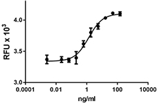
Compare Data Across All Formats
This data display is provided for general comparisons between formats.
Your actual data may vary due to variations in samples, target cells, instruments and their settings, staining conditions, and other factors.
If you need assistance with selecting the best format contact our expert technical support team.


Follow Us