 Login / Register


- Clone
- 7H8.2C12 (See other available formats)
- Regulatory Status
- RUO
- Other Names
- Cyt c
- Isotype
- Mouse IgG2b, κ
- Ave. Rating
- Submit a Review
- Product Citations
- publications

-

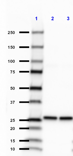
Total lysates (15 µg protein) from HeLa (Human), Raw264.7 (Mouse), UMR106 (Rat) and CHO (Hamster) were resolved by electrophoresis (4-20% Tris-glycine gel), transferred to nitrocellulose, and probed with 1:1000 purified anti-Cytochrome C antibody, clone 7H8.2C12. Proteins were visualized using chemiluminescence detection by incubation with HRP Goat anti-Mouse secondary antibody (Cat. No. 405306, 1:3000 dilution). Non specific band was marked with “*”. Direct-Blot™ HRP anti-β-actin was used as a loading control (Cat. No. 643807, 1:8000 dilution). -

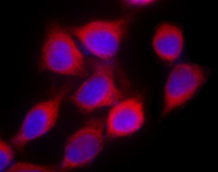
Hela cells were pre-incubated with 400nM MitoSpy™ Green FM at 37°C for 30 minutes and then fixed with 4% paraformaldehyde (PFA) for 15 minutes, permeabilized with 0.5% Triton X-100 for 3 minutes, and blocked with 5% FBS for 60 minutes. The cells were intracellularly stained with either (A) 3 µg/ml anti-mouse IgG1, κ, or (B) anti-cytochrome C antibody (clone 7H8.2C12), and incubated overnight at 4°C followed by 27micro;g/ml (1:250) Alexa Fluor® 594 (Red) goat anti-mouse IgG for one hour at room temperature. Nuclei were counterstained with DAPI. The image was captured with a 60X objective. Exposure time for (A) is 1/30, and (B) is 1/60. -

HeLa cells were treated with 400 nM MitoSpy™ Green FM (Green, Cat. No. 424805) for 30 minutes, fixed with 4% paraformaldehyde (PFA) for fifteen minutes, permeabilized with 0.5% Triton X-100 for three minutes, and blocked with 5% FBS for 60 minutes. Then the cells were intracellularly stained with purified anti-Cytochrome c antibody (clone 7H8.2C12) overnight at 4°C followed by Alexa Fluor® 594 (Red) goat anti-mouse IgG for one hour at room temperature (Cat. No. 405326, 1:250 dilution, 2 µg/ml). Nuclei were counterstained with DAPI (blue, Cat. No. 422801). The image was captured with a 60X objective. The image was captured with a 60X objective using KEYENCE BZ-X700 fluorescence microscope. Exposure time (in seconds) is 1/20. -

HeLa cells were treated with 400 nM MitoSpy™ Red CMXRos (Red, Cat. No. 424801) for 30 minutes, fixed with 4% paraformaldehyde (PFA) for fifteen minutes, permeabilized with 0.5% Triton X-100 for three minutes, and blocked with 5% FBS for 60 minutes. Then the cells were intracellularly stained with purified anti-Cytochrome c antibody (clone 7H8.2C12) overnight at 4°C followed by Alexa Fluor® 488 (Green) goat anti-mouse IgG for one hour at room temperature (Cat. No. 405319, 1:250 dilution, 2 µg/ml). Nuclei were counterstained with DAPI (Blue, Cat. No. 422801). The image was captured with a 60X objective. The image was captured with a 60X objective using KEYENCE BZ-X700 fluorescence microscope. Exposure time (in seconds) is 1/20.
Cytochrome c is a 15 kD protein found in the mitochondrial intermembrane space with a heme-binding domain. Cytochrome c is a component of the electron transport chain; the heme group transfers electrons from cytochrome b-c1 complex to cytochrome oxidase complex. Cytochrome c initiates apoptosis by release to cytoplasm and binding Apaf-1 which activates procaspase 9. Cytochrome c interacts with the cytochrome b-c1 complex, cytochrome oxidase complex, heme, Apaf-1, and Caspase 9 proteins. The 7H8.2C12 monoclonal antibody recognizes cytochrome-c from most species and has been shown to be useful for Western blotting.
Product DetailsProduct Details
- Verified Reactivity
- Human, Mouse, Rat, Hamster
- Reported Reactivity
- Other species
- Antibody Type
- Monoclonal
- Host Species
- Mouse
- Immunogen
- Horse cyt c-OVA
- Formulation
- This antibody is provided in phosphate-buffered solution, pH 7.2, containing 0.09% sodium azide.
- Preparation
- The antibody was purified by affinity chromatography.
- Concentration
- Lot-specific (to obtain lot-specific concentration and expiration, please enter the lot number in our Certificate of Analysis online tool.)
- Storage & Handling
- Upon receipt, store undiluted between 2°C and 8°C.
- Application
-
WB - Quality tested
ICC - Verified - Recommended Usage
-
Each lot of this antibody is quality control tested by Western blotting. Western blotting, suggested working dilution(s): Use 10 µl antibody per 5 ml antibody dilution buffer for each mini-gel. For immunocytochemistry, a concentration range of 1.0 - 3.0 µg/ml (1:150-1:500 dilution) is recommended. It is recommended that the reagent be titrated for optimal performance for each application.
-
Application References
(PubMed link indicates BioLegend citation) -
- Jemmerson R, et al. 1991. Eur. J. Immunol. 21:143. (WB)
- Semenkova L, et al. 2003. Eur. J. Biochem. 270:4388. (WB)
- Product Citations
-
- RRID
-
AB_2090157 (BioLegend Cat. No. 612503)
AB_2292697 (BioLegend Cat. No. 612504)
Antigen Details
- Structure
- Heme binding domain; 15 kD
- Distribution
-
Mitochondrial intermembrane space
- Function
- Component of electron transport chain; heme group transfers electrons from cytochrome b-c1 complex to cytochrome oxidase complex. Initiates apoptosis by release to cytoplasm and binding Apaf-1 which activates procaspase 9
- Interaction
- Cytochrome b-c1 complex, cytochrome oxidase complex, heme, Apaf-1, Casp9
- Biology Area
- Apoptosis/Tumor Suppressors/Cell Death, Cell Biology, Mitochondrial Function, Neuroscience, Neuroscience Cell Markers
- Molecular Family
- Mitochondrial Markers
- Antigen References
-
1. Liu X, et al. 1996. Cell 86:147.
2. Li P, et al. 1997. Cell 91:479.
3. Zhang Z, et al. 2003. Gene 312:61.
4. Ferguson H, et al. 2003. J. Biol. Chem. 278:45793. - Gene ID
- 54205 View all products for this Gene ID
- UniProt
- View information about Cytochrome c on UniProt.org

Related FAQs
Other Formats
View All Cytochrome c Reagents Request Custom Conjugation| Description | Clone | Applications |
|---|---|---|
| Purified anti-Cytochrome c | 7H8.2C12 | WB,ICC |
Customers Also Purchased
Compare Data Across All Formats
This data display is provided for general comparisons between formats.
Your actual data may vary due to variations in samples, target cells, instruments and their settings, staining conditions, and other factors.
If you need assistance with selecting the best format contact our expert technical support team.


Follow Us