Propidium Iodide Solution

Pricing & Availability
Regulatory Status
RUO
Other Names
PI
Ave. Rating
Submit a Review
Product Citations
publications
Cat # Size Price Quantity Check Availability Save
421301 2 mL 44€
Check Availability


Need larger quantities of this item?
Request Bulk Quote
Description

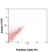
Propidium iodide (PI) is a fluorescent dye that binds to DNA. When excited by 488nm laser light, it can be detected with in the PE/Texas Red® channel with a bandpass filter 610/10. It is commonly used in evaluation of cell viability or DNA content in cell cycle analysis by flow cytometry.

Product Details
Technical data sheet

Product Details

Formulation
Phosphate-buffered saline, pH 7.2, containing 0.09% sodium azide.
Concentration
0.5 mg/ml
Storage & Handling
The solution should be stored undiluted between 2°C and 8°C, and protect from light. Caution: This solution contains hazardous material; handle with care.
Application

FC - Quality tested

Recommended Usage

The suggested use of this solution for viability staining is 10 µl per million cells in 0.5 ml/test, and incubate for 15 minutes at 4 °C before analysis. For Cell Cycle analysis, please see our Propidium Iodide Cell Cycle Staining Protocol. Caution: This solution is toxigenic and mutagenic; handle with care.

Excitation Laser
Blue Laser (488 nm)
Application Notes

Propidium Iodide Solution can be used in evaluation of apoptosis, cell viability and cell cycle analysis by flow cytometry.

Application References

(PubMed link indicates BioLegend citation)
  1. Vermes I, et al.1995. J. Immunol. Methods 184:39.
  2. Darzynkiewicz Z, et al. 1992. Cytometry 13(8):795.
  3. Douglas RS, et al. 1995. J. Immunol. Methods 188:219.
  4. Sakimoto I, et al. 2006. Cancer Research 66:2287.PubMed
  5. Liu H, et al. 2012. Int J Biochem Cell Biol. 45:408. PubMed
  6. Juel HB, et al. 2013. PLoS One. 8:64619. PubMed
  7. Ren Q, et al. 2013. PLoS One. 8:74732. PubMed
  8. Zhao W, et al. 2013. Clin Immunol. 149:119. PubMed
  9. Beggs KM, et al. 2014. Toxicol Sci. 137:91. PubMed
  10. Ewald SE, et al. 2014. Infect Immun. 82:460. PubMed
  11. Tsou WI, et al. 2014. J Biol Chem. 289:25750. PubMed
  12. Kong S, et al. 2014. J Immunol. 193:5515. PubMed
Product Citations
  1. Liu H, et al. 2012. Int J Biochem Cell Biol. 45:408. PubMed
  2. Ewald S, et al. 2014. Infect Immun . 82:460. PubMed
  3. Guo H, et al. 2016. Sci Rep. 6:37559. PubMed
  4. Smith A, et al. 2017. Sci Rep. 7:41872. PubMed
  5. Li W, et al. 2019. Aging (Albany NY). 0.99375. PubMed
  6. Chan J, et al. 2017. Oncogene. 10.1038/onc.2017.319. PubMed
  7. Uroda T, et al. 2019. Mol Cell. 75:982. PubMed
  8. Lindqvist B, et al. 2022. PLoS Pathog. 18:e1010555. PubMed
  9. Neumann CKA, et al. 2020. Mol Metab. 34:136. PubMed
  10. Hamel Y, et al. 2021. Cell Rep Med. 2:100370. PubMed
  11. Castellanos CA, et al. 2021. Sci Immunol. 6:eabh0707. PubMed
  12. Leeth C, et al. 2021. Animals (Basel). 11:. PubMed
  13. Chen M, et al. 2021. Cancers (Basel). 13:. PubMed
  14. McNeal AS, et al. 2021. Elife. 10:. PubMed
  15. Ren Q, et al. 2013. PLoS One. 8:74732. PubMed
  16. He Y, et al. 2020. Nat Commun. 11:1996. PubMed
  17. Fukushima T, et al. 2019. Cell Rep. 29:4144. PubMed
  18. Yoshioka H, et al. 2021. Front Cell Dev Biol. 9:621045. PubMed
  19. Yang Y, et al. 2021. Metabolism. 123:154863. PubMed
  20. Sun X, et al. 2022. JCI Insight. 7: . PubMed
  21. Coray M, et al. 2022. Int J Mol Sci. 23:. PubMed
  22. Schrier S, et al. 2016. Sci Rep. 6:34942. PubMed
  23. O'Neill K, et al. 2016. Genes Dev. 30: 973-988. PubMed
  24. Sapanidou V, et al. 2015. Theriogenology. . PubMed
  25. Kong S, et al. 2014. J Immunol. 193:5515. PubMed
  26. Zeng H, et al. 2018. Cancer Cell. 34:56:00. PubMed
  27. Lei W, et al. 2020. Sci Rep. 10:7004. PubMed
  28. Wang Y, et al. 2019. Cell. 179:1144. PubMed
  29. Zhang M, et al. 2017. Oncol Lett. 3.069444444. PubMed
  30. Zhang W, et al. 2021. J Clin Invest. 131:. PubMed
  31. Ambrosi TH, et al. 2021. Elife. 10: . PubMed
  32. Feng L, et al. 2016. J Cell Sci. 129: 2202 - 2212. PubMed
  33. Lange N, et al. 2016. PLoS One. 11: 0160843. PubMed
  34. Jordan PM, et al. 2020. Cell Rep. 33:108247. PubMed
  35. Chen C, et al. 2019. Cell Rep. 29:4200. PubMed
  36. Liu L, et al. 2019. Cell Prolif. :e12718. PubMed
  37. Zou PA, et al. 2021. Oncol Lett. 22:648. PubMed
  38. Masuda T, et al. 2022. iScience. 25:104781. PubMed
  39. Reighard SD, et al. 2020. Cell Rep Med. :1. PubMed
  40. Zhang S, et al. 2020. Front Immunol. 1.757638889. PubMed
  41. Xu B, et al. 2016. Leukemia. 10.1038/leu.2016.297. PubMed
  42. Liu Y, et al. 2017. Acta Biomater. 10.1016/j.actbio.2017.08.021. PubMed
  43. Domenis R, et al. 2017. PLoS One. 10.1371/journal.pone.0169932. PubMed
  44. Walter F, et al. 2020. PLoS One. 15:e0239369. PubMed
  45. Yang M, et al. 2021. Med Sci Monit. 27:e930083. PubMed
  46. Zhang X, et al. 2020. Eur J Med Chem. 112186:192. PubMed
  47. Mensen A, et al. 2015. Biol Blood Marrow Transplant. Available online 10 August 2015. PubMed
  48. Zhao W, et al. 2013. Clin Immunol. 149:119. PubMed
  49. Liu H, et al. 2020. Cell Death Dis. 0.767361111. PubMed
  50. Li L, et al. 2018. Oncol Lett. 4.184722222. PubMed
  51. Jung E, et al. 2017. J Neurosci. 37:6837. PubMed
  52. Basso-Ricci L, et al. 2017. Cytometry A. 10.1002/cyto.a.23148. PubMed
  53. Matsuda K, et al. 2021. Cell Rep Methods. 1:100122. PubMed
  54. Pang K, et al. 2022. Cell Death Dis. 13:169. PubMed
  55. Tang X, et al. 2021. Transl Cancer Res. 10:2120. PubMed
  56. Li Y, et al. 2016. Int J Lower Extrem Wounds. 10.1177/1534734616649485. PubMed
  57. Hu D, et al. 2020. Proc Natl Acad Sci U S A. 117:30639. PubMed
  58. Li Q, et al. 2020. Acta Biochim Biophys Sin (Shanghai). 52:1131. PubMed
  59. Moribe F, et al. 2021. PLoS One. 16:e0237554. PubMed
  60. Litzenburger UM, et al. 2017. Genome Biol. 18:15. PubMed
  61. Shanahan MT, et al. 2021. Am J Physiol Gastrointest Liver Physiol. 321:G668. PubMed
  62. Wang J, et al. 2022. J Clin Invest. :. PubMed
  63. Trudu M, et al. 2022. Front Oncol. 12:968208. PubMed
  64. L Chang, Y Li, D Kaplan 2016. Sci Rep. 6:36862. PubMed
  65. Perl AL, et al. 2019. J Biol Chem. 294:17043. PubMed
  66. Diaz Osterman CJ, et al. 2019. Elife. 8:e47327. PubMed
  67. Hosokawa T, et al. 2017. J Immunol. 10.4049/jimmunol.1700157. PubMed
  68. El-Hafeez A, et al. 2017. Cytotechnology. 10.1007/s10616-017-0124-1. PubMed
  69. Xue G, et al. 2021. Nat Biomed Eng. 5:1306. PubMed
  70. Lasrado N, et al. 2022. iScience. 25:103865. PubMed
  71. Tsou W, et al. 2014. J Biol Chem. 289:25750. PubMed
  72. Oubaha M, et al. 2016. Sci Transl Med. 8: 362ra144. PubMed
  73. Polk A, et al. 2016. Clin Cancer Res. 22(24):6118-6128. PubMed
  74. Harro CM, et al. 2020. J Clin Invest. . PubMed
  75. Imura Y, et al. 2020. JCI Insight. :5. PubMed
  76. Suzuki S, et al. 2021. STAR Protocols. 2:100683. PubMed
  77. Vickman R, et al. 2016. Mol Cancer Res. 14: 776 - 786. PubMed
  78. El-Hafeez A, et al. 2017. Cytotechnology. 10.1007/s10616-017-0116-1. PubMed
  79. Xie M, et al. 2017. BMC Cancer . 10.1186/s12885-017-3291-8. PubMed
  80. Pal P, et al. 2021. J Med Chem. 64:14230. PubMed
  81. Hedemann N, et al. 2021. Cancers (Basel). 13:. PubMed
  82. Xie B, et al. 2021. J Immunol. 206:1515. PubMed
  83. M H, et al. 2016. Cell Transplant. 25: 811-27. PubMed
  84. Woo MS, et al. 2021. J Exp Med. :218. PubMed
  85. Huang K, et al. 2018. Biol Open. 7:bio029470. PubMed
  86. Wang H, et al. 2019. Nat Commun. 4.388194444. PubMed
  87. Matsubara T, et al. 2022. J Exp Med. 219:. PubMed
  88. Hughes K, et al. 2017. Open Bio. 10.1098/rsob.160155. PubMed
  89. Resch U, et al. 2016. MBio. 7: e00207-16. PubMed
  90. Bao W, et al. 2016. Sci Rep. 6:22579. PubMed
  91. Liu X, et al. 2019. Mol Cancer Ther. 18:706. PubMed
  92. Yang X, et al. 2017. J Control Release. 10.1016/j.jconrel.2017.07.014. PubMed
  93. Viereckl MJ, et al. 2022. Biomolecules. 12:. PubMed
  94. Deng Y, et al. 2021. Nat Commun. 12:7041. PubMed
  95. Beggs K, et al. 2014. Toxicol Sci. 137:91. PubMed
  96. Wang C, et al. 2017. Drug Deliv. 10.1080/10717544.2017.1293195. PubMed
  97. Nilsri N, et al. 2021. Sci Rep. 5255:11. PubMed
  98. Bjørnstad R, et al. 2019. Mol Cancer Ther. 1.14375. PubMed
  99. Ghura H, et al. 2021. Neoplasia. 23:837. PubMed
  100. He Y, et al. 2020. J Hematol Oncol. 0.607638889. PubMed
  101. Widodo N, et al. 2022. F1000Res. 11:169. PubMed
  102. Tu Z, et al. 2022. Cell Rep. 40:111072. PubMed
  103. Bensberg M, et al. 2021. Proc Natl Acad Sci U S A. 118:. PubMed
  104. Namas R, et al. 2016. Lupus Sci Med. 3: e000148. PubMed
  105. Chen Y, et al. 2016. J Biol Chem. 291: 13216 - 13228. PubMed
  106. Deng BB, et al. 2020. Cell Biol Int. 44:1890. PubMed
  107. Siddhartha V, et al. 2017. Artif Cells Nanomed Biotechnol . 10.1080/21691401.2017.1313267. PubMed
  108. Viseshakul N, et al. 2017. Parasitol Res. 10.1007/s00436-017-5589-5. PubMed
  109. Chmielewski M and Abken H 2017. Cell Rep.. 10.1016/j.celrep.2017.11.063. PubMed
  110. Sakimoto I, et al. 2006. Cancer Res. 66:2287. PubMed
  111. Goel S, et al. 2021. Nat Commun. 12:5325. PubMed
  112. Szeto C, et al. 2022. Nat Commun. 13:4951. PubMed
  113. Liu Q, et al. 2016. Cell Death Dis. 1.93125. PubMed
  114. Mahmoudi M, et al. 2016. Sci Rep. 6:26960. PubMed
  115. Juel H, et al. 2013. PLoS One. 8:64619. PubMed
  116. Popa S, et al. 2020. Wellcome Open Res. 0.249305556. PubMed
  117. Vulin M, et al. 2022. Oncogene. :. PubMed

Antigen Details

Biology Area
Cell Biology, Cell Cycle/DNA Replication, Neuroscience
Gene ID
NA

Related FAQs

Why is washing not recommended after the addition 7-AAD or PI addition when assessing viability?

These dyes bind in equilibrium with DNA. Therefore, external dye concentration must be maintained during analysis and the dye should not be washed out.

Go To Top Version: 2    Revision Date: 10.08.2015

For Research Use Only. Not for diagnostic or therapeutic use.


This product is supplied subject to the terms and conditions, including the limited license, located at www.biolegend.com/terms) ("Terms") and may be used only as provided in the Terms. Without limiting the foregoing, BioLegend products may not be used for any Commercial Purpose as defined in the Terms, resold in any form, used in manufacturing, or reverse engineered, sequenced, or otherwise studied or used to learn its design or composition without express written approval of BioLegend. Regardless of the information given in this document, user is solely responsible for determining any license requirements necessary for user’s intended use and assumes all risk and liability arising from use of the product. BioLegend is not responsible for patent infringement or any other risks or liabilities whatsoever resulting from the use of its products.


BioLegend, the BioLegend logo, and all other trademarks are property of BioLegend, Inc. or their respective owners, and all rights are reserved.


8999 BioLegend Way, San Diego, CA 92121 www.biolegend.com
Toll-Free Phone: 1-877-Bio-Legend (246-5343) Phone: (858) 768-5800 Fax: (877) 455-9587

ProductsHere

Login / Register
Remember me
Forgot your password? Reset password?
Create an Account