 Login / Register


- Clone
- hC3aRZ8 (See other available formats)
- Regulatory Status
- RUO
- Other Names
- C3a anaphylatoxin chemotactic receptor, C3a-R, AZ3B, HNFAG09
- Isotype
- Mouse IgG2b, κ
- Ave. Rating
- Submit a Review
- Product Citations
- publications

-

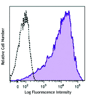
Human peripheral blood granulocytes were stained with human C3AR (clone hC3aRZ8) PE/Cyanine7 (filled histogram) or mouse IgG2b, ? PE/Cyanine7 isotype contrl (open histogram).
| Cat # | Size | Price | Quantity Check Availability | Save | ||
|---|---|---|---|---|---|---|
| 345807 | 25 tests | 147€ | ||||
| 345808 | 100 tests | 306€ | ||||
C3AR is a single chain protein of 482 aa, with seven membrane-spanning regions and a molecular weight of 54 Kd. C3AR is expressed by neutrophils, eosinophils, mast cells, monocytes, dendritic cells and there are reports of C3AR expression in non-immune cells such as hematopoietic and mesenchymal stem cells, epithelial and endothelial cells. C3AR is coupled to heterotrimeric G proteins and after C3a binding, the signal transduced, results in chemotaxis, granule enzyme release and superoxide anion production.
Product DetailsProduct Details
- Verified Reactivity
- Human
- Antibody Type
- Monoclonal
- Host Species
- Mouse
- Immunogen
- hC3aR transfected cells
- Formulation
- Phosphate-buffered solution, pH 7.2, containing 0.09% sodium azide and BSA (origin USA)
- Preparation
- The antibody was purified by affinity chromatography and conjugated with PE/Cyanine7 under optimal conditions.
- Concentration
- Lot-specific (to obtain lot-specific concentration and expiration, please enter the lot number in our Certificate of Analysis online tool.)
- Storage & Handling
- The antibody solution should be stored undiluted between 2°C and 8°C, and protected from prolonged exposure to light. Do not freeze.
- Application
-
FC - Quality tested
- Recommended Usage
-
Each lot of this antibody is quality control tested by immunofluorescent staining with flow cytometric analysis. For flow cytometric staining, the suggested use of this reagent is 5 µl per million cells in 100 µl staining volume or 5 µl per 100 µl of whole blood.
- Excitation Laser
-
Blue Laser (488 nm)
Green Laser (532 nm)/Yellow-Green Laser (561 nm)
- Application Notes
-
Additional reported applications (for the relevant formats) include: immunohistochemical staining of acetone-fixed frozen sections2.
-
Application References
(PubMed link indicates BioLegend citation) -
- Soruri A, et al. 2002. J. Immunol. 170:3306 (FC)
- Kiafard Z, et al. 2007 Immunobiology. 212:129 (FC, IHC)
- RRID
-
AB_2810549 (BioLegend Cat. No. 345807)
AB_2810550 (BioLegend Cat. No. 345808)
Antigen Details
- Structure
- 482 aa, single chain protein with seven membrane-spanning regions, 54 kD
- Distribution
-
Neutrophils, eosinophils, mast cells, monocytes, dendritic cells, hematopoietic and mesenchymal stem cells, endothelial cells
- Function
- After C3a binding, the C3AR signaling results in chemotaxis, granule enzyme release and superoxide anion production.
- Interaction
- G proteins
- Bioactivity
- Chemotaxis, degranulation
- Cell Type
- Dendritic cells, Endothelial cells, Eosinophils, Mast cells, Mesenchymal Stem Cells, Monocytes, Neutrophils
- Biology Area
- Cell Biology, Costimulatory Molecules, Immunology, Innate Immunity, Neuroinflammation, Neuroscience, Stem Cells
- Molecular Family
- Cytokine/Chemokine Receptors
- Antigen References
-
1. Wysoczynski M et al. 2009. Leukemia. 23:1455
2. Schraufstatter IU. et al. 2009. J. Immunol. 182:3827
3. Honczarenko M et al. 2005. J. Immunol. 175:3698
4. Reca R et al. 2003. Blood 101:3784 - Gene ID
- 719 View all products for this Gene ID
- UniProt
- View information about C3AR on UniProt.org


Related FAQs
Other Formats
View All C3aR Reagents Request Custom Conjugation| Description | Clone | Applications |
|---|---|---|
| Purified anti-human C3AR | hC3aRZ8 | FC,IHC-F |
| PE anti-human C3AR | hC3aRZ8 | FC |
| APC anti-human C3AR | hC3aRZ8 | FC |
| PE/Cyanine7 anti-human C3AR | hC3aRZ8 | FC |
| TotalSeq™-A1038 anti-human C3AR | hC3aRZ8 | PG |
| TotalSeq™-C1038 anti-human C3AR | hC3aRZ8 | PG |
Customers Also Purchased
Compare Data Across All Formats
This data display is provided for general comparisons between formats.
Your actual data may vary due to variations in samples, target cells, instruments and their settings, staining conditions, and other factors.
If you need assistance with selecting the best format contact our expert technical support team.
-
Purified anti-human C3AR

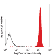
Human peripheral blood granulocytes stained with hC3aRZ8 PE ... -
PE anti-human C3AR
Human peripheral blood granulocytes stained with hC3aRZ8 PE ... -
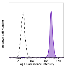
APC anti-human C3AR

Human peripheral blood granulocytes were stained with C3aR (... -
PE/Cyanine7 anti-human C3AR

Human peripheral blood granulocytes were stained with human ... -
TotalSeq™-A1038 anti-human C3AR
-
TotalSeq™-C1038 anti-human C3AR


Follow Us