 Login / Register


- Clone
- JES6-5H4 (See other available formats)
- Regulatory Status
- RUO
- Other Names
- Interleukin-2, T cell growth factor (TCGF), Eosinophil differentiation factor (EDF), Killer cell helper factor (KHF), Macrophage-activating factor for cytotoxicity I (MAF-C I), Thymocyte differentiation factor (TDF)
- Isotype
- Rat IgG2b, κ
- Ave. Rating
- Submit a Review
- Product Citations
- publications

-

PMA+ionomycin-stimulated C57BL/6 mouse splenocytes intracellular stained with with CD3 (17A2) APC and JES6-5H4 PE
| Cat # | Size | Price | Quantity Check Availability | Save | ||
|---|---|---|---|---|---|---|
| 503807 | 25 µg | 67€ | ||||
| 503808 | 100 µg | 176€ | ||||
IL-2 is a potent lymphoid cell growth factor which exerts its biological activity primarily on T cells. Additionally, IL-2 has been found to stimulate growth and differentiation of B cells, NK cells, LAK cells, monocytes, and oligodendrocytes.
Product DetailsProduct Details
- Verified Reactivity
- Mouse
- Antibody Type
- Monoclonal
- Host Species
- Rat
- Immunogen
- E. coli-expressed, recombinant mouse IL-2
- Formulation
- Phosphate-buffered solution, pH 7.2, containing 0.09% sodium azide.
- Preparation
- The antibody was purified by affinity chromatography, and conjugated with PE under optimal conditions.
- Concentration
- 0.2 mg/ml
- Storage & Handling
- The antibody solution should be stored undiluted between 2°C and 8°C, and protected from prolonged exposure to light. Do not freeze.
- Application
-
ICFC - Quality tested
- Recommended Usage
-
Each lot of this antibody is quality control tested by intracellular immunofluorescent staining with flow cytometric analysis. For flow cytometric staining, the suggested use of this reagent is ≤1.0 µg per million cells in 100 µl volume. It is recommended that the reagent be titrated for optimal performance for each application.
- Excitation Laser
-
Blue Laser (488 nm)
Green Laser (532 nm)/Yellow-Green Laser (561 nm)
- Application Notes
-
ELISA Detection1-3 or ELISPOT Detection4-6: The biotinylated JES6-5H4 antibody is useful as a detection antibody for a sandwich ELISA or ELISPOT assay, when used in conjunction with the purified JES6-1A12 antibody (Cat. Nos. 503701 & 503702) as capture antibody and recombinant mouse IL-2 (Cat. No. 575409) as the standard.
Flow Cytometry8-10: The fluorochrome-labeled JES6-5H4 antibody is useful for intracellular immunofluorescent staining and flow cytometric analysis to identify IL-2 -producing cells within mixed cell populations.
Neutralization1,7: The Ultra-LEAF™ purified antibody (Endotoxin in vivo and in vitro (Cat. No. 503845-503850)) is recommended for neutralization.
Additional reported applications (for the relevant formats) include: immunoprecipitation1, immunohistochemical staining of paraformaldehyde-fixed, saponin-treated frozen tissue sections2, in vivo capture7, and immunocytochemistry.
Note: For testing mouse IL-2 in serum, plasma or supernatant, BioLegend's ELISA MAX™ Sets (Cat. No. 431001 & 431004) are specially developed and recommended. -
Application References
(PubMed link indicates BioLegend citation) -
- Abrams J, et al. 1992. Immunol. Rev. 127:5.
- Sander B, et al. 1993. J. Immunol. Meth. 166:201.
- Abrams J. 1995. Curr. Prot. Immunol. John Wiley and Sons New York. Unit 6.20.
- Klinman D, et al. 1994. Curr. Prot. Immunol. John Wiley and Sons New York. Unit 6.19.
- Mo X, et al. 1995. J. Virol. 69:1288.
- Karulin A, et al. 2000. J. Immunol. 164:1862.
- Finkelman F, et al. 2003. Curr. Prot. Immunol. John Wiley & Sons New York. Unit 6.28.
- Ko SY, et al. 2005. J. Immunol. 175:3309. PubMed
- Kang SS and Allen PM. 2005. J. Immunol. 174:5382.
- Lawson BR, et al. 2007. J. Immunol. 178:5366.
- Product Citations
-
- RRID
-
AB_315301 (BioLegend Cat. No. 503807)
AB_315302 (BioLegend Cat. No. 503808)
Antigen Details
- Structure
- Cytokine; 15-30 kD (Mammalian)
- Bioactivity
- Proliferation of T lymphocytes, B cells, anti-inflammatory, hematopoiesis, tumor surveillance
- Cell Sources
- T cells
- Cell Targets
- T cells, B cells, NK cells, LAK cells, monocytes, macrophages, oligodendrocytes
- Receptors
- High affinity heterotrimer of IL-2Rα/β/γ, intermediate affinity homodimer IL-2Rα (CD25; p55; Tac) and heterodimer IL-2Rβ (CD122)/γ; γ-subunit (CD132) in common with IL-4R, IL-7R, IL-13R, IL-15R
- Cell Type
- Tregs
- Biology Area
- Immunology
- Molecular Family
- Cytokines/Chemokines
- Antigen References
-
1. Fitzgerald K, et al. Eds. 2001. The Cytokine FactsBook. Academic Press San Diego.
2. Taniguchi T, et al. 1993. Cell 73:5.
3. Nistico G. 1993. Prog. Neurobiol. 40:463.
4. Waldmann T, et al. 1993. Ann. NY Acad. Sci. 685:603. - Regulation
- Upregulated by NFAT; downregulated by TCF-8, CIF (colostrum inhibitory factor)
- Gene ID
- 16183 View all products for this Gene ID
- UniProt
- View information about IL-2 on UniProt.org
Related Pages & Pathways
Pathways


Related FAQs
- What type of PE do you use in your conjugates?
- We use R-PE in our conjugates.
Other Formats
View All IL-2 Reagents Request Custom ConjugationCustomers Also Purchased
Compare Data Across All Formats
This data display is provided for general comparisons between formats.
Your actual data may vary due to variations in samples, target cells, instruments and their settings, staining conditions, and other factors.
If you need assistance with selecting the best format contact our expert technical support team.
-
APC anti-mouse IL-2

PMA+ionomycin stimulated C57BL/6 mouse splenocytes (6 hours)... -
Biotin anti-mouse IL-2

-
FITC anti-mouse IL-2

PMA-ionomycin-stimulated Balb/c mouse splenocytes intracellu... -
PE anti-mouse IL-2

PMA+ionomycin-stimulated C57BL/6 mouse splenocytes intracell... -
Purified anti-mouse IL-2
-
Alexa Fluor® 488 anti-mouse IL-2

PMA+ionomycin-stimulated (6 hours) Balb/c mouse splenocytes ... -
Alexa Fluor® 647 anti-mouse IL-2

PMA/Ionomycin stimulatd (6 hrs) BALB/c splenocytes intracell... -
Alexa Fluor® 700 anti-mouse IL-2

PMA+ionomycin-stimulated C57BL/6 mouse splenocytes (6 hours)... -
Pacific Blue™ anti-mouse IL-2

PMA+ionomycin-stimulated C57BL/6 mouse splenocytes (6 hours)... -
PerCP/Cyanine5.5 anti-mouse IL-2

C57BL/6 mouse splenocytes stimulated with PMA + Ionomycin (6... -
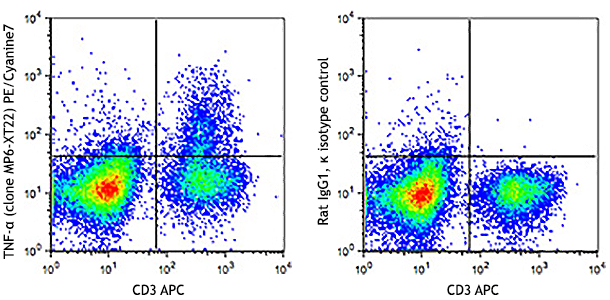
PE/Cyanine5 anti-mouse IL-2

PMA/Ionomycin-stimulated C57BL/6 mouse splenocytes (6 hours)... -
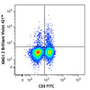
Brilliant Violet 421™ anti-mouse IL-2

C57BL/6 mouse splenocytes were stimulated with PMA + Ionomyc... 
-
Brilliant Violet 605™ anti-mouse IL-2

PMA+ionomycin-stimulated C57BL/6 mouse splenocytes (in the p... -
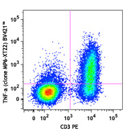
PE/Cyanine7 anti-mouse IL-2

PMA+ionomycin-stimulated C57BL/6 mouse splenocytes (in the p... -
Brilliant Violet 510™ anti-mouse IL-2

PMA+ionomycin-stimulated (6 hours) C57BL/6 mouse splenocytes... 
-
Purified anti-mouse IL-2 (Maxpar® Ready)

C57BL/6 mouse splenocytes were incubated for 18 hours in med... -
Brilliant Violet 711™ anti-mouse IL-2

PMA+Ionomycin-stimulated (6 hours) C57BL/6 mouse splenocytes... 
-
PE/Dazzle™ 594 anti-mouse IL-2

C57BL/6 mouse splenocytes were stimulated with PMA + Ionomyc... 
-
APC/Fire™ 750 anti-mouse IL-2

C57BL/6 mouse splenocytes were stimulated with Cell Activati... -
Brilliant Violet 785™ anti-mouse IL-2

C57BL/6 mouse splenocytes were stimulated with Cell Activati... -
Spark Red™ 718 anti-mouse IL-2

C57BL/6 mouse splenocytes were stimulated with the Cell Acti...


Follow Us