 Login / Register


- Clone
- AA10 (See other available formats)
- Regulatory Status
- RUO
- Other Names
- β-3 Tubulin, Neuronal class III beta-tubulin, β3-tub
- Isotype
- Mouse IgG2a, κ
- Ave. Rating
- Submit a Review
- Product Citations
- publications

-

Human lung adenocarcinoma cell line A549 was fixed with 1% paraformaldehyde (PFA) for ten minutes, permeabilized with 0.5 % Triton X-100 for ten minutes, and blocked with 5% FBS for 30 minutes. Then, the tissue was stained with 2.5 µg/ml of Brilliant Violet 421™ anti-Tubulin Beta 3 (TUBB3) (clone AA10, blue) overnight at 4°C. Nuclei were counterstained with DRAQ5™ (red). The image was captured with a 40X objective. -

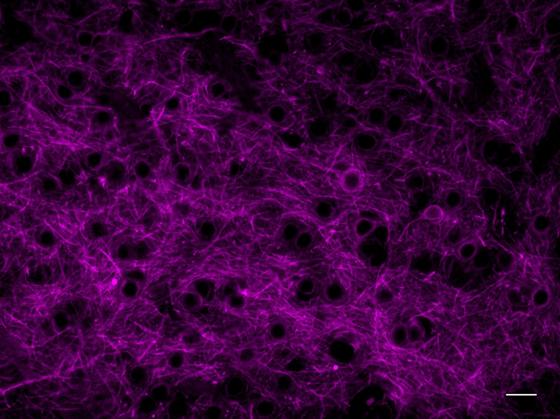
C57BL/6 mouse frozen brain (cerebellum) tissue was fixed with 4% paraformaldehyde (PFA) for ten minutes, permeabilized with 0.5 % Triton X-100 for ten minutes, and blocked with 5% FBS for one hour. Then, the tissue was stained with 2.5 µg/ml of Brilliant Violet 421™ anti-Tubulin Beta 3 (TUBB3) (clone AA10, blue) and 5 µg/ml of Alexa Fluor® 488 anti-GFAP (clone 2E1.E9, green) in 5% FBS, overnight at 4°C. Nuclei were counterstained with DRAQ5™ (red). The image was captured with a 10X objective.
| Cat # | Size | Price | Quantity Check Availability | Save | ||
|---|---|---|---|---|---|---|
| 657412 | 100 µg | 348€ | ||||
Tubulin is the main component of microtubules. In adults, tubulin beta 3 (TUBB3) is primarily expressed in neurons and is commonly used as a neuronal marker. It plays an important role in neuronal cell proliferation and differentiation. Mutations in this gene cause congenital fibrosis of the type 3 extraocular muscles. Tubulin beta 3 (TUBB3) is also found in a wide range of tumors. Studies indicate that it is a predictive and prognostic marker in various tumors.
Product DetailsProduct Details
- Verified Reactivity
- Mouse, Rat, Human
- Antibody Type
- Monoclonal
- Host Species
- Mouse
- Immunogen
- Fusion protein
- Formulation
- Phosphate-buffered solution, pH 7.2, containing 0.09% sodium azide and BSA (origin USA).
- Preparation
- The antibody was purified by affinity chromatography and conjugated with Brilliant Violet 421™ under optimal conditions.
- Concentration
- 0.2 mg/ml
- Storage & Handling
- The antibody solution should be stored undiluted between 2°C and 8°C, and protected from prolonged exposure to light. Do not freeze.
- Application
-
IHC-F - Quality tested
ICC - VerifiedSB - Reported in the literature, not verified in house
- Recommended Usage
-
Each lot of this antibody is quality control tested by immunohistochemical staining on frozen tissue sections. For immunohistochemical staining on frozen tissue sections, the suggested use of this reagent is 1.25 - 5.0 µg/ml. For immunocytochemistry, a concentration range of 1.25 - 5.0 μg/ml is recommended. It is recommended that the reagent be titrated for optimal performance for each application.
Brilliant Violet 421™ excites at 405 nm and emits at 421 nm. The standard bandpass filter 450/50 nm is recommended for detection. Brilliant Violet 421™ is a trademark of Sirigen Group Ltd.
Learn more about Brilliant Violet™.
This product is subject to proprietary rights of Sirigen Inc. and is made and sold under license from Sirigen Inc. The purchase of this product conveys to the buyer a non-transferable right to use the purchased product for research purposes only. This product may not be resold or incorporated in any manner into another product for resale. Any use for therapeutics or diagnostics is strictly prohibited. This product is covered by U.S. Patent(s), pending patent applications and foreign equivalents. - Excitation Laser
-
Violet Laser (405 nm)
- Application Notes
-
Additional reported application (for relevant formats) include: spatial biology (IBEX)1,2.
- Additional Product Notes
-
Iterative Bleaching Extended multi-pleXity (IBEX) is a fluorescent imaging technique capable of highly-multiplexed spatial analysis. The method relies on cyclical bleaching of panels of fluorescent antibodies in order to image and analyze many markers over multiple cycles of staining, imaging, and, bleaching. It is a community-developed open-access method developed by the Center for Advanced Tissue Imaging (CAT-I) in the National Institute of Allergy and Infectious Diseases (NIAID, NIH).
-
Application References
(PubMed link indicates BioLegend citation) - RRID
-
AB_2632699 (BioLegend Cat. No. 657412)
Antigen Details
- Structure
- 450 amino acids with predicted molecular weight of 50 kD
- Distribution
-
Cytosol
- Function
- Plays important roles in neuronal cell proliferation and differentiation
- Interaction
- Alpha tubulin, kinesin and dynein
- Cell Type
- Mature Neurons
- Biology Area
- Cell Biology, Cell Cycle/DNA Replication, Cell Motility/Cytoskeleton/Structure, Immunology, Neuroscience, Neuroscience Cell Markers
- Molecular Family
- Microtubules
- Antigen References
-
1. Katsetos CD, et al. 2003. J. Child Neurol. 18:851.
2. Mobarakeh ZT, et al. 2012. Cell Biol. Int. Rep. (2010) 19:e00015.
3. Locher H, et al. 2013. Differentiation. 85:173.
4. Karki R, et al. 2013. Expert Opin. Ther. Targets. 17:461.
5. Mariani M, et al. 2011. Curr. Mol. Med. 11:726.
6. Koh Y, et al. 2009. Ann. Oncol. 20:1414. - Gene ID
- 10381 View all products for this Gene ID
- UniProt
- View information about Tubulin beta-3 on UniProt.org


Related FAQs
- What is the F/P ratio range of our BV421™ format antibody reagents?
-
It is lot-specific. On average it ranges between 2-4.
- If an antibody clone has been previously successfully used in IBEX in one fluorescent format, will other antibody formats work as well?
-
It’s likely that other fluorophore conjugates to the same antibody clone will also be compatible with IBEX using the same sample fixation procedure. Ultimately a directly conjugated antibody’s utility in fluorescent imaging and IBEX may be specific to the sample and microscope being used in the experiment. Some antibody clone conjugates may perform better than others due to performance differences in non-specific binding, fluorophore brightness, and other biochemical properties unique to that conjugate.
- Will antibodies my lab is already using for fluorescent or chromogenic IHC work in IBEX?
-
Fundamentally, IBEX as a technique that works much in the same way as single antibody panels or single marker IF/IHC. If you’re already successfully using an antibody clone on a sample of interest, it is likely that clone will have utility in IBEX. It is expected some optimization and testing of different antibody fluorophore conjugates will be required to find a suitable format; however, legacy microscopy techniques like chromogenic IHC on fixed or frozen tissue is an excellent place to start looking for useful antibodies.
- Are other fluorophores compatible with IBEX?
-
Over 18 fluorescent formats have been screened for use in IBEX, however, it is likely that other fluorophores are able to be rapidly bleached in IBEX. If a fluorophore format is already suitable for your imaging platform it can be tested for compatibility in IBEX.
- The same antibody works in one tissue type but not another. What is happening?
-
Differences in tissue properties may impact both the ability of an antibody to bind its target specifically and impact the ability of a specific fluorophore conjugate to overcome the background fluorescent signal in a given tissue. Secondary stains, as well as testing multiple fluorescent conjugates of the same clone, may help to troubleshoot challenging targets or tissues. Using a reference control tissue may also give confidence in the specificity of your staining.
- How can I be sure the staining I’m seeing in my tissue is real?
-
In general, best practices for validating an antibody in traditional chromogenic or fluorescent IHC are applicable to IBEX. Please reference the Nature Methods review on antibody based multiplexed imaging for resources on validating antibodies for IBEX.
Other Formats
View All Tubulin β-3 (TUBB3) Reagents Request Custom Conjugation| Description | Clone | Applications |
|---|---|---|
| Purified anti-Tubulin Beta 3 (TUBB3) | AA10 | WB,ICC |
| Alexa Fluor® 488 anti-Tubulin Beta 3 (TUBB3) | AA10 | ICFC,IHC-F,3D IHC |
| Alexa Fluor® 647 anti-Tubulin Beta 3 (TUBB3) | AA10 | ICFC,IHC-F,3D IHC,SB |
| Alexa Fluor® 594 anti-Tubulin Beta 3 (TUBB3) | AA10 | IHC-F,ICC,IHC-P,3D IHC |
| Direct-Blot™ HRP anti-Tubulin Beta 3 (TUBB3) | AA10 | WB |
| Brilliant Violet 421™ anti-Tubulin Beta 3 (TUBB3) | AA10 | IHC-F,ICC,SB |
Customers Also Purchased
Compare Data Across All Formats
This data display is provided for general comparisons between formats.
Your actual data may vary due to variations in samples, target cells, instruments and their settings, staining conditions, and other factors.
If you need assistance with selecting the best format contact our expert technical support team.
-
Purified anti-Tubulin Beta 3 (TUBB3)


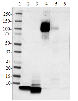
Mouse brain (lane 1), mouse liver (lane 2), N-tera2 (lane 3)... 
Whole cell lysates (15 µg protein) from HEL (negative contro... 
NTERA‐2 cells were fixed with 4% paraformaldehyde (PFA) for ... -
Alexa Fluor® 488 anti-Tubulin Beta 3 (TUBB3)

Human lung adenocarcinoma cell line A549 was treated with Bi... 
C57BL/6 mouse frozen brain tissue was fixed with 4% paraform... 
Dissected C57/B6 mouse small intestine was immersed in 4% p... 
Formalin-fixed, 300 micron-thick mouse kidney section was bl... -
Alexa Fluor® 647 anti-Tubulin Beta 3 (TUBB3)

Human lung adenocarcinoma cell line A549 was treated with Fi... 
C57BL/6 mouse frozen brain tissue was fixed with 4% paraform... Formalin-fixed, 300 micron-thick mouse large intestine secti... -
Alexa Fluor® 594 anti-Tubulin Beta 3 (TUBB3)

Day-three cultured postnatal C57BL/6 mouse brain cells were ... 
C57BL/6 mouse frozen brain tissue was fixed with 4% paraform... 
C57BL/6 mouse frozen brain tissue was fixed with 4% paraform... 
Human paraffin-embedded cerebellum tissue slices were prepar... 
Human paraffin-embedded cerebellum tissue slices were prepar... Formalin-fixed, 300 micron-thick mouse brain (cerebellum) se... -
Direct-Blot™ HRP anti-Tubulin Beta 3 (TUBB3)

10 µg of total protein extract from HeLa cells was resolved ... -
Brilliant Violet 421™ anti-Tubulin Beta 3 (TUBB3)

Human lung adenocarcinoma cell line A549 was fixed with 1% p... 
C57BL/6 mouse frozen brain (cerebellum) tissue was fixed wit...


Follow Us