 Login / Register


- Clone
- HK5.3 (See other available formats)
- Regulatory Status
- RUO
- Other Names
- ICOS ligand, ICOSL, B7 homolog 2, B7-H2, B7H2, B7RP1, B7h, ICOS-L
- Isotype
- Rat IgG2a, κ
- Ave. Rating
- Submit a Review
- Product Citations
- publications

-

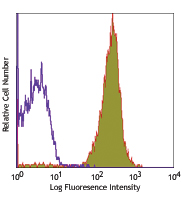
C57Bl/6 mouse splenocytes stained with biotinylated HK5.3, followed by Sav-PE
| Cat # | Size | Price | Quantity Check Availability | Save | ||
|---|---|---|---|---|---|---|
| 107403 | 50 µg | 90€ | ||||
CD275 is a 40 kD protein, also known as ICOS ligand, B7-RP1, B7-H2, and B7h. It is a member of the immunoglobulin receptor superfamily expressed on splenic B cells, dendritic cells, and macrophages. The expression of this antigen is increased by TNF-α stimulation and is highly upregulated by local inflammation. B7RP1 binds to ICOS and co-stimulates T cell and B cell responses. Two isoforms of this protein have been reported to occur as a result of alternative splicing. These isoforms show differential tissue distribution.
Product DetailsProduct Details
- Verified Reactivity
- Mouse
- Antibody Type
- Monoclonal
- Host Species
- Rat
- Immunogen
- Mouse B7-RP1 transfected cell line
- Formulation
- Phosphate-buffered solution, pH 7.2, containing 0.09% sodium azide.
- Preparation
- The antibody was purified by affinity chromatography, and conjugated with biotin under optimal conditions.
- Concentration
- 0.5 mg/ml
- Storage & Handling
- The antibody solution should be stored undiluted between 2°C and 8°C. Do not freeze.
- Application
-
FC - Quality tested
- Recommended Usage
-
Each lot of this antibody is quality control tested by immunofluorescent staining with flow cytometric analysis. For flow cytometric staining, the suggested use of this reagent is ≤1.0 µg per million cells in 100 µl volume. It is recommended that the reagent be titrated for optimal performance for each application.
- Application Notes
-
Additional reported applications (for the relevant formats) include: in vitro and in vivo blocking of ligand binding1,2. For most successful immunofluorescent staining results, it may be important to maximize signal over background by using a relatively bright fluorochrome-antibody conjugate (Cat. No. 107405) or by using a high sensitivity, three-layer staining technique (e.g., including a biotinylated antibody (Cat. No. 107404) or biotinylated anti-rat IgG second step (Cat. No. 405402), followed by SAv-PE (Cat. No. 405204)). The LEAF™ purified antibody (Endotoxin <0.1 EU/µg, Azide-Free, 0.2 µm filtered) is recommended for functional assays (Cat. No. 107408). For in vivo studies or highly sensitive assays, we recommend Ultra-LEAF™ purified antibody (Cat. No. 107410) with a lower endotoxin limit than standard LEAF™ purified antibodies (Endotoxin <0.01 EU/µg).
-
Application References
(PubMed link indicates BioLegend citation) - Product Citations
-
- RRID
-
AB_345259 (BioLegend Cat. No. 107403)
Antigen Details
- Structure
- B7 Immunoglobulin superfamily, approximately 40 kD
- Distribution
-
Isoform 1 has highest expression in lymphoid tissues, splenic B-cells, T-cells, dendritic cells and macrophages; lower levels in many non-lymphoid tissues. The expression of isoform 2 is restricted to heart, spleen, and kidney.
- Function
- Binds to ICOS. Co-stimulates T cell responses including proliferation and cytokine secretion. Stimulates B cell proliferation and differentiation to plasma cells.
- Ligand/Receptor
- ICOS
- Cell Type
- B cells, Dendritic cells, Lymphocytes, Macrophages, T cells
- Biology Area
- Costimulatory Molecules, Immunology
- Molecular Family
- CD Molecules
- Antigen References
-
1. Yoshinaga SK, et al. 1999. Nature 402:827.
2. Ling V, et al. 2000. J. Immunol. 164:1653.
3. Ling V, et al. 2001. J. Immunol. 164:1653. - Gene ID
- 50723 View all products for this Gene ID
- UniProt
- View information about CD275 on UniProt.org
Related Pages & Pathways
Pathways


Related FAQs
- How many biotin molecules are per antibody structure?
- We don't routinely measure the number of biotins with our antibody products but the number of biotin molecules range from 3-6 molecules per antibody.
Other Formats
View All CD275 Reagents Request Custom Conjugation| Description | Clone | Applications |
|---|---|---|
| Biotin anti-mouse CD275 (B7-H2, B7-RP1, ICOS Ligand) | HK5.3 | FC |
| PE anti-mouse CD275 (B7-H2, B7-RP1, ICOS Ligand) | HK5.3 | FC |
| Ultra-LEAF™ Purified anti-mouse CD275 (B7-H2, B7-RP1, ICOS Ligand) | HK5.3 | FC,Block |
Customers Also Purchased
Compare Data Across All Formats
This data display is provided for general comparisons between formats.
Your actual data may vary due to variations in samples, target cells, instruments and their settings, staining conditions, and other factors.
If you need assistance with selecting the best format contact our expert technical support team.
-
Biotin anti-mouse CD275 (B7-H2, B7-RP1, ICOS Ligand)

C57Bl/6 mouse splenocytes stained with biotinylated HK5.3, f... -
PE anti-mouse CD275 (B7-H2, B7-RP1, ICOS Ligand)

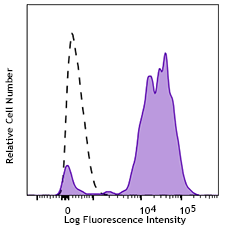
C57Bl/6 mouse splenocytes stained with CD275 (clone HK5.3) P... -
Ultra-LEAF™ Purified anti-mouse CD275 (B7-H2, B7-RP1, ICOS Ligand)
C57Bl/6 mouse splenocytes stained with CD275 (clone HK5.3) P...


Follow Us