 Login / Register


- Clone
- TY25 (See other available formats)
- Regulatory Status
- RUO
- Other Names
- PD-L2, PDL2, B7DC, CD273
- Isotype
- Rat IgG2a, κ
- Ave. Rating
- Submit a Review
- Product Citations
- publications

-

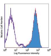
mB7-DC transfected cells stained with biotinylated TY25, followed by Sav-PE
| Cat # | Size | Price | Quantity Check Availability | Save | ||
|---|---|---|---|---|---|---|
| 107203 | 50 µg | 90€ | ||||
B7-DC is also called programmed death ligand 2 (PDL2). It has recently been clustered as CD273. This ligand is a 42 kD member of the immunoglobulin receptor superfamily expressed on a subset of dendritic cells, liver and a small subset of macrophages as well as a few transformed cell lines. CD273 has been reported to be stimulatory on dendritic cells when cross-linked and to inhibit T cell activation upon engaging the PD-1 receptor. CD273 has also been reported to bind to an alternative receptor and to mediate T cell activation through such non-PD1 mediated interactions. The TY25 antibody has been reported to be useful for blocking PD-1 mediated interactions.
Product DetailsProduct Details
- Verified Reactivity
- Mouse
- Antibody Type
- Monoclonal
- Host Species
- Rat
- Immunogen
- Mouse B7-DC transfected cell line
- Formulation
- Phosphate-buffered solution, pH 7.2, containing 0.09% sodium azide.
- Preparation
- The antibody was purified by affinity chromatography, and conjugated with biotin under optimal conditions.
- Concentration
- 0.5 mg/ml
- Storage & Handling
- The antibody solution should be stored undiluted between 2°C and 8°C. Do not freeze.
- Application
-
FC - Quality tested
- Recommended Usage
-
Each lot of this antibody is quality control tested by immunofluorescent staining with flow cytometric analysis. For flow cytometric staining, the suggested use of this reagent is ≤ 0.25 µg per 106 cells in 100 µl volume. It is recommended that the reagent be titrated for optimal performance for each application.
- Application Notes
-
Additional reported applications (for the relevant formats) include: immunoprecipitation1, Western blotting1,6, blocking2,4,5 of PD-1 mediated interactions, and immunohistochemistry of acetone-fixed frozen sections3. The Ultra-LEAF™ purified antibody (Endotoxin < 0.01 EU/µg, Azide-Free, 0.2 µm filtered) is recommended for functional assays (Cat. Nos. 107221-107226).
-
Application References
(PubMed link indicates BioLegend citation) -
- Yamazaki T, et al. 2002. J. Immunol. 169:5538. (FC, IP, WB)
- Ansari MJI, et al. 2003. J. Exp. Med. 198:63. (Block)
- Salama AD, et al. 2003. J. Exp. Med. 198:71. (IHC)
- Matsumoto K, et al. 2004. J. Immunol. 172:2530. (FC, Block)
- Yamazaki T, et al. 2005. J. Immunol. 175:1586. (Block)
- Meng Q, et al. 2006. Invest Opthalmol Vis Sci. 47:444. (WB) PubMed
- del Rio ML, et al. 2011. Transpl. Int. 24:501. (FC) PubMed
- Murakami R, et al. 2013. PLoS One. 8:73270. PubMed
- Product Citations
-
- RRID
-
AB_345251 (BioLegend Cat. No. 107203)
Antigen Details
- Structure
- B7 Immunoglobulin superfamily, 42 kD
- Distribution
-
Dendritic cells, liver, few transformed cell lines, subset of macrophages; not expressed on lymphoid tissues
- Function
- Binds to PD-1 and alternative receptor; ligation on DC stimulates, inhibits T cell responses via PD-1 binding, stimulates T cells via alternative receptor binding and promotes tumor immunity
- Ligand/Receptor
- PD-1, uncharacterized receptor
- Cell Type
- Dendritic cells, Macrophages
- Biology Area
- Costimulatory Molecules, Immunology
- Molecular Family
- CD Molecules, Immune Checkpoint Receptors
- Antigen References
-
1. Shin T, et al. 2003. J. Exp. Med. 198:31.
2. Liu X, et al. 2003. J. Exp. Med. 197:1721.
3. Carreno BM, et al. 2002. Annu. Rev. Immunol. 20:29. - Gene ID
- 58205 View all products for this Gene ID
- UniProt
- View information about CD273 on UniProt.org
Related Pages & Pathways
Pathways


Related FAQs
- How many biotin molecules are per antibody structure?
- We don't routinely measure the number of biotins with our antibody products but the number of biotin molecules range from 3-6 molecules per antibody.
Other Formats
View All CD273 Reagents Request Custom Conjugation| Description | Clone | Applications |
|---|---|---|
| Purified anti-mouse CD273 (B7-DC, PD-L2) | TY25 | FC,IHC-F,IP,WB |
| Biotin anti-mouse CD273 (B7-DC, PD-L2) | TY25 | FC |
| PE anti-mouse CD273 (B7-DC, PD-L2) | TY25 | FC |
| APC anti-mouse CD273 (B7-DC, PD-L2) | TY25 | FC |
| Brilliant Violet 421™ anti-mouse CD273 (B7-DC, PD-L2) | TY25 | FC |
| PE/Cyanine7 anti-mouse CD273 (B7-DC, PD-L2) | TY25 | FC |
| PerCP/Cyanine5.5 anti-mouse CD273 (B7-DC, PD-L2) | TY25 | FC |
| PE/Dazzle™ 594 anti-mouse CD273 (B7-DC, PD-L2) | TY25 | FC |
| Ultra-LEAF™ Purified anti-mouse CD273 (B7-DC, PD-L2) | TY25 | FC,IHC-F,IP,WB |
| TotalSeq™-A0914 anti-mouse CD273 (B7-DC, PD-L2) | TY25 | PG |
| TotalSeq™-C0914 anti-mouse CD273 (B7-DC, PD-L2) | TY25 | PG |
Customers Also Purchased
Compare Data Across All Formats
This data display is provided for general comparisons between formats.
Your actual data may vary due to variations in samples, target cells, instruments and their settings, staining conditions, and other factors.
If you need assistance with selecting the best format contact our expert technical support team.
-
Purified anti-mouse CD273 (B7-DC, PD-L2)

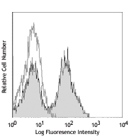
mB7-DC transfected cells stained with purified TY25, followe... -
Biotin anti-mouse CD273 (B7-DC, PD-L2)

mB7-DC transfected cells stained with biotinylated TY25, fol... -
PE anti-mouse CD273 (B7-DC, PD-L2)

mB7-DC transfected cells stained with TY25 PE -
APC anti-mouse CD273 (B7-DC, PD-L2)

Mouse B7-DC transfected BHK cells were stained with CD273 (c... -
Brilliant Violet 421™ anti-mouse CD273 (B7-DC, PD-L2)

Mouse B7-DC transfected BHK cells were stained with CD273 (c... -
PE/Cyanine7 anti-mouse CD273 (B7-DC, PD-L2)

Mouse B7-DC transfected BHK cells were stained with CD273 (c... -
PerCP/Cyanine5.5 anti-mouse CD273 (B7-DC, PD-L2)

Mouse B7-DC transfected BHK cells were stained with CD273 (B... -
PE/Dazzle™ 594 anti-mouse CD273 (B7-DC, PD-L2)

Mouse B7-DC transfected BHK cells were stained with CD273 (c... -
Ultra-LEAF™ Purified anti-mouse CD273 (B7-DC, PD-L2)

mB7-DC transfected cells stained with LEAF™ purified TY25, f... -
TotalSeq™-A0914 anti-mouse CD273 (B7-DC, PD-L2)
-
TotalSeq™-C0914 anti-mouse CD273 (B7-DC, PD-L2)


Follow Us