 Login / Register


- Clone
- PE001 (See other available formats)
- Regulatory Status
- RUO
- Other Names
- PE
- Isotype
- Mouse IgG1, κ
- Ave. Rating
- Submit a Review
- Product Citations
- publications

-

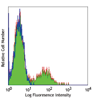
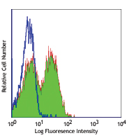
C57BL/6 thymocytes stained with CD4 PE and anti-phycoerythrin (left) APC or mouse IgG1, κ isotype control (right) APC.
| Cat # | Size | Price | Quantity Check Availability | Save | ||
|---|---|---|---|---|---|---|
| 408107 | 25 µg | 63€ | ||||
| 408108 | 100 µg | 167€ | ||||
The PE001 antibody reacts with both phycoerythrin (PE) alone and PE conjugated with antibody or streptavidin, as well as PE tandem dyes such as PE/Cy5, PE/Cy5.5 and PE/Cy7. When PE001 antibody is used in a three-step staining procedure or separation of PE-antibody labeled cells, it does not quench PE fluorescence or react with tested mouse and human cells. The PE001 antibody can be used for separation of PE-antibody labeled cells or as a secondary step to amplify PE conjugated primary antibody signals (i.e. biotinylated PE001 as secondary step, followed by Streptavidin-PE).
Product DetailsProduct Details
- Antibody Type
- Monoclonal
- Host Species
- Mouse
- Immunogen
- Phycoerythrin
- Formulation
- Phosphate-buffered solution, pH 7.2, containing 0.09% sodium azide.
- Preparation
- The antibody was purified by affinity chromatography and conjugated with APC under optimal conditions.
- Concentration
- 0.2 mg/ml
- Storage & Handling
- The antibody solution should be stored undiluted between 2°C and 8°C, and protected from prolonged exposure to light. Do not freeze.
- Application
-
FC - Quality tested
- Recommended Usage
-
Each lot of this antibody is quality control tested by immunofluorescent staining with flow cytometric analysis. For flow cytometric staining, the suggested use of this reagent is ≤ 0.5 µg per million cells in 100 µl volume. It is recommended that the reagent be titrated for optimal performance for each application.
- Excitation Laser
-
Red Laser (633 nm)
-
Application References
(PubMed link indicates BioLegend citation) -
- Taylor RT, et al. 2007. J. Immunol. 178:5659.
- Product Citations
-
- RRID
-
AB_2814387 (BioLegend Cat. No. 408107)
AB_2814388 (BioLegend Cat. No. 408108)
Antigen Details
- Gene ID
- NA
- UniProt
- View information about Phycoerythrin on UniProt.org


Related FAQs
- What type of PE do you use in your conjugates?
- We use R-PE in our conjugates.
Other Formats
View All Phycoerythrin Reagents Request Custom Conjugation| Description | Clone | Applications |
|---|---|---|
| Purified anti-phycoerythrin (PE) | PE001 | FC |
| Biotin anti-phycoerythrin (PE) | PE001 | FC |
| Purified anti-phycoerythrin (PE) (Maxpar® Ready) | PE001 | FC,CyTOF® |
| APC anti-phycoerythrin (PE) | PE001 | FC |
| TotalSeq™-A0911 anti-phycoerythrin (PE) | PE001 | PG |
| TotalSeq™-C0911 anti-phycoerythrin (PE) | PE001 | PG |
| TotalSeq™-B0911 anti-phycoerythrin (PE) | PE001 | PG |
Customers Also Purchased
Compare Data Across All Formats
This data display is provided for general comparisons between formats.
Your actual data may vary due to variations in samples, target cells, instruments and their settings, staining conditions, and other factors.
If you need assistance with selecting the best format contact our expert technical support team.
-
Purified anti-phycoerythrin (PE)

C57BL/6 splenocytes stained with RM4-5 PE and purified PE001... -
Biotin anti-phycoerythrin (PE)

C57BL/6 thymocytes stained with RM4-5 PE only (top) or RM4-5... 
C57BL/6 thymocytes stained with RM4-5 PE only (top) or RM4-5... -
Purified anti-phycoerythrin (PE) (Maxpar® Ready)

Human PBMC stained with PE-anti-CD3 (UCHT1) then detected wi... -
APC anti-phycoerythrin (PE)

C57BL/6 thymocytes stained with CD4 PE and anti-phycoerythri... -
TotalSeq™-A0911 anti-phycoerythrin (PE)
-
TotalSeq™-C0911 anti-phycoerythrin (PE)
-
TotalSeq™-B0911 anti-phycoerythrin (PE)


Follow Us