 Login / Register


- Clone
- 1F11 (See other available formats)
- Regulatory Status
- RUO
- Other Names
- P2X7 receptor (P2X7R), P2RX7
- Isotype
- Rat IgG2b, κ
- Ave. Rating
- Submit a Review
- Product Citations
- publications

-

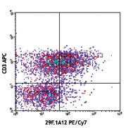
C57BL/6 splenocytes were stained with CD4 Pacific Blue™ and P2X7R (clone 1F11) APC (top) or rat IgG2b, κ APC isotype control (bottom). -

| Cat # | Size | Price | Quantity Check Availability | Save | ||
|---|---|---|---|---|---|---|
| 148705 | 25 µg | 124€ | ||||
| 148706 | 100 µg | 266€ | ||||
P2X7R, also known as P2X7 receptor, belongs to the family of ligand-gated ion channel receptors. It is expressed on T cells, B cells, macrophages, and microglia. The receptor opens in the presence of extracellular ATP or NAD, leading to intracellular calcium mobilization. P2X7R activation requires higher concentrations of ATP compared to other P2X receptors. Longer stimulation results in larger pores, allowing passage of larger molecules. Activation of these molecules also leads to mitochondrial and cytoskeletal changes as well as IL-1β maturation and release. Ligation of P2X7 receptor can lead to membrane blebbing and cell death.
Product DetailsProduct Details
- Verified Reactivity
- Mouse
- Antibody Type
- Monoclonal
- Host Species
- Rat
- Immunogen
- Murine colon mast cells
- Formulation
- Phosphate-buffered solution, pH 7.2, containing 0.09% sodium azide.
- Preparation
- The antibody was purified by affinity chromatography and conjugated with APC under optimal conditions.
- Concentration
- 0.2 mg/ml
- Storage & Handling
- The antibody solution should be stored undiluted between 2°C and 8°C, and protected from prolonged exposure to light. Do not freeze.
- Application
-
FC - Quality tested
- Recommended Usage
-
Each lot of this antibody is quality control tested by immunofluorescent staining with flow cytometric analysis. For flow cytometric staining, the suggested use of this reagent is ≤0.25 µg per million cells in 100 µl volume. It is recommended that the reagent be titrated for optimal performance for each application.
- Excitation Laser
-
Red Laser (633 nm)
- Application Notes
-
Additional reported applications for the relevant formats include: immunoprecipitation1, Western blotting1, immunohistochemistry1, in vivo inhibition of intestinal inflammation and mast cell activation1.
-
Application References
(PubMed link indicates BioLegend citation) -
- Kurashima Y, et al. 2012. Nat. Commun. 3:1034. (FC, IP, WB, IHC, FA)
- Product Citations
-
- RRID
-
AB_2650953 (BioLegend Cat. No. 148705)
AB_2650954 (BioLegend Cat. No. 148706)
Antigen Details
- Structure
- Ligand-gated ion channel, trimer - two transmembrane regions and one cytosolic region.
- Distribution
-
Immune cells, including mast cells, microglia, macrophages, T cells, and B cells.
- Function
- Ion channel activated by extracellular ATP.
- Ligand/Receptor
- ATP, NAD.
- Cell Type
- B cells, Macrophages, Mast cells, T cells, Tregs
- Biology Area
- Apoptosis/Tumor Suppressors/Cell Death, Cell Biology, Immunology, Innate Immunity, Neuroscience, Neuroscience Cell Markers
- Antigen References
-
1. Surprenant A, et al. 1996. Science. 272:735.
2. Chused TM, et al. 1996. J. Immunol. 157:1371.
3. Gargett CE, et al. 1997. Br. J. Pharmacol. 122:911.
4. Kawamura H, et al. 2006. J. Immunol. 176:2152.
5. Pelegrin P. 2011. Br. J. Pharmacol. 163:908.
6. Bartlett R, et al. 2014. Pharmacol. Rev. 66:638.
7. Alves LA, et al. 2014. Biochim. Biophys. Acta. 1838:2578. - Gene ID
- 18439 View all products for this Gene ID
- UniProt
- View information about P2X7R on UniProt.org


Related FAQs
Other Formats
View All P2X7R Reagents Request Custom Conjugation| Description | Clone | Applications |
|---|---|---|
| Purified anti-mouse P2X7R | 1F11 | FC,IP,WB,IHC |
| APC anti-mouse P2X7R | 1F11 | FC |
| PE anti-mouse P2X7R | 1F11 | FC |
| PE/Cyanine7 anti-mouse P2X7R | 1F11 | FC |
| PerCP/Cyanine5.5 anti-mouse P2X7R | 1F11 | FC |
| TotalSeq™-A0824 anti-mouse P2X7R | 1F11 | PG |
| TotalSeq™-C0824 anti-mouse P2X7R | 1F11 | PG |
| TotalSeq™-B0824 anti-mouse P2X7R Antibody | 1F11 | PG |
Customers Also Purchased

Compare Data Across All Formats
This data display is provided for general comparisons between formats.
Your actual data may vary due to variations in samples, target cells, instruments and their settings, staining conditions, and other factors.
If you need assistance with selecting the best format contact our expert technical support team.
-
Purified anti-mouse P2X7R

C57BL/6 splenocytes were stained with CD4 FITC and purified ... 
-
APC anti-mouse P2X7R

C57BL/6 splenocytes were stained with CD4 Pacific Blue™ and ... 
-
PE anti-mouse P2X7R

C57BL/6 splenocytes were stained with CD4 (clone RM4-5) Pac... -
PE/Cyanine7 anti-mouse P2X7R

C57BL/6 splenocytes were stained with CD4 APC and P2X7R (clo... -
PerCP/Cyanine5.5 anti-mouse P2X7R

C57BL/6 splenocytes were stained with CD4 FITC and P2X7R (cl... -
TotalSeq™-A0824 anti-mouse P2X7R
-
TotalSeq™-C0824 anti-mouse P2X7R
-
TotalSeq™-B0824 anti-mouse P2X7R Antibody


Follow Us