 Login / Register


- Clone
- O91D3 (See other available formats)
- Regulatory Status
- RUO
- Other Names
- CTRCT30, HEL 113, Epididymis Luminal Protein 113
- Isotype
- Mouse IgG2a, κ
- Ave. Rating
- Submit a Review
- Product Citations
- publications

-

Total cell lysates (15 µg total protein) from Daudi (negative control), PC-3, Jurkat and NIH/3T3 cells were resolved by 4-12% Bis-Tris gel electrophoresis, transferred to a nitrocellulose membrane, and probed with 0.25 µg/mL (1:2000 dilution) of Purified anti-Vimentin Antibody, clone O91D3, overnight at 4°C. Proteins were visualized by chemiluminescence detection using HRP goat anti-mouse IgG Antibody (Cat. No. 405306) at a 1:3000 dilution. Direct-Blot™ HRP anti-GAPDH Antibody (Cat. No. 607904) was used as a loading control at a 1:50000 dilution (lower). Lane M: Molecular Weight marker. Predicted expression data was obtained from Human Protein Atlas. -

HeLa cells were stained with purified anti-Vimentin (clone O91D3) antibody, followed by staining with Alexa Fluor® 594 conjugated goat anti-mouse IgG antibody (red). Actin filaments were labeled with Alexa Fluor® 647 Phalloidin (green). Nuclei were counterstained with DAPI (blue). The image was captured with a 40X objective. -

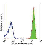
Jurkat cells (filled histogram, positive control) and Daudi cells (open histogram, negative control) were fixed with Fixation Buffer (Cat. No. 420801), permeabilized using True-Phos™ Perm Buffer (Cat. No. 425401), and intracellularly stained with 0.25 µg/test of purified anti-Vimentin antibody (clone O91D3) followed by PE goat anti-mouse IgG antibody (Cat. No. 405307). -

IHC staining of anti-Vimentin antibody (clone O91D3) on formalin-fixed paraffin-embedded human kidney (A) and brain (B) tissues. Following antigen retrieval using Sodium Citrate H.I.E.R. (Cat. No. 928502), the tissues were incubated with 5 µg/mL of the anti-Vimentin antibody overnight at 4°C. BioLegend’s Ultra Streptavidin HRP Kit (Multi-Species, DAB, Cat. No. 929501) was used for detection followed by hematoxylin counterstaining, according to the protocol provided. The images were captured with a 40X objective. Scale bar: 50 μm. -

IHC staining of anti-Vimentin antibody (clone O91D3) on frozen human spleen (A) and brain (B) tissues. Following fixation with Fixation Buffer (Cat. No. 420801) and permeabilization with 0.5% Triton X-100, the tissue sections were incubated with 5.0 µg/mL of anti-Vimentin antibody overnight at 4°C followed by incubation with Alexa Fluor® 594 Goat anti-mouse IgG antibody (Cat. No. 405326) for 2-hours at room temperature. Nuclei were counterstained with DAPI (Cat. No. 422801) and the slides were mounted with ProLong™ Gold Antifade Mountant. These images were captured with a 40X objective. Scale bar: 50μm. -

SeqIF™ (sequential immunofluorescence) staining on COMET™ of Purified anti-Vimentin (clone O91D3, yellow) on formalin-fixed paraffin-embedded human pancreatic carcinoma at 0.33 µg/mL. Alexa Fluor™ Plus 647 Goat anti-Mouse IgG antibody (Lunaphore, Cat. No. DR647MS) was used as a secondary antibody. Nuclei were counterstained with DAPI (blue). Tissue underwent an all-in-one dewaxing and antigen retrieval preprocessing.
| Cat # | Size | Price | Quantity Check Availability | Save | ||
|---|---|---|---|---|---|---|
| 677801 | 25 µg | 81€ | ||||
| 677802 | 100 µg | 212€ | ||||
Vimentin are class-III intermediate filaments found in various non-epithelial cells, especially mesenchymal cells. Vimentin is a widely expressed and highly conserved 54 kD protein that is constitutively expressed in mesenchymal cells, endothelial cells lining blood vessels, renal tubular cells, macrophages, neutrophils, fibroblasts, and leukocytes1,2. Vimentin is used as a marker of mesenchymal cells to distinguish them from epithelial cells3. Increased vimentin expression is frequently used as an EMT marker in cancer4. Autoantibodies to vimentin are commonly found in patients with autoimmune diseases such as Lupus5 and rheumatoid arthritis6, and also found after transplantation7.
Product DetailsProduct Details
- Verified Reactivity
- Human
- Antibody Type
- Monoclonal
- Host Species
- Mouse
- Immunogen
- Full length human vimentin produced in E. coli.
- Formulation
- Phosphate-buffered solution, pH 7.2, containing 0.09% sodium azide.
- Preparation
- The antibody was purified by affinity chromatography.
- Concentration
- 0.5 mg/mL
- Storage & Handling
- The antibody solution should be stored undiluted between 2°C and 8°C.
- Application
-
WB - Quality tested
ICC, ICFC, IHC-P, IHC-F - Verified
SB - Community verified - Recommended Usage
-
Each lot of this antibody is quality control tested by Western blotting. For Western blotting, the suggested use of this reagent is 0.25 - 2.5 µg per mL. For immunocytochemistry, a concentration range of 1.0 - 5.0 µg/mL is recommended. For flow cytometric staining, the suggested use of this reagent is ≤ 0.25 µg per million cells in 100 µL volume. For immunohistochemistry on formalin-fixed paraffin-embedded tissue sections, a concentration range of 1.0 - 5.0 µg/mL is suggested. For immunohistochemistry on frozen tissue sections, a concentration range of 1.0 - 10.0 µg/mL is suggested. It is recommended that the reagent be titrated for optimal performance for each application.
- Application Notes
-
While this clone recognizes mouse Vimentin, we do not recommend its usage for western blot due to poor affinity of the antibody for the protein. Additional reported applications for the relevant formats include: spatial biology (IBEX)1,2.
- Additional Product Notes
-
For the use of this antibody in spatial biology applications, we have partnered with Lunaphore Technologies for demonstration of our antibodies on the COMET™. The COMET™ platform is an automated, end-to-end spatial biology solution developed for rapid and flexible multiplex tissue profiling. More information on the COMET™ and a complete list of our antibodies that have been demonstrated on the COMET™ can be found here.
-
Application References
(PubMed link indicates BioLegend citation) - Product Citations
-
- RRID
-
AB_2565911 (BioLegend Cat. No. 677801)
AB_2565982 (BioLegend Cat. No. 677802)
Antigen Details
- Structure
- 466 amino acids with a predicted molecular weight of approximately 54 kD.
- Distribution
-
Cytoplasm.
- Function
- Vimentins are class-III intermediate filaments found in various non-epithelial cells, especially mesenchymal cells. Vimentin is attached to the nucleus, endoplasmic reticulum, and mitochondria, either laterally or terminally.
- Interaction
- HCV core protein, LGSN, SYNM, PLEC, SLC6A4, STK33, LARP6, RAB8B, TOR1A, TOR1AIP1, and BCAS3.
- Cell Type
- B cells, Mesenchymal Stem Cells, Neural Stem Cells, Neutrophils
- Biology Area
- Cell Adhesion, Cell Biology, Cell Motility/Cytoskeleton/Structure, Immunology, Neuroscience, Neuroscience Cell Markers, Stem Cells
- Molecular Family
- Intermediate Filaments
- Antigen References
-
1. Kidd ME, et al. 2014. Am. J. Respir. Cell Mol. Biol. 50:1.
2. Fuchs E, et al. 1994. Annu. Rev. Biochem. 63:345.
3. Zeisberg M, et al. 2009. J. Clin. Invest. 119:1429.
4. Scanlon CS, et al. 2013. J. Dent. Res. 92:114.
5. Thebault S, et al. 2002. J. Immunol. 169:4046.
6. Vossenaar ER, et al. 2004. Arthritis Res. Ther. 6:R142.
7. Rose ML. 2013. Hum. Immunol. 74:1459. - Gene ID
- 7431 View all products for this Gene ID
- UniProt
- View information about Vimentin on UniProt.org

Related FAQs
- If an antibody clone has been previously successfully used in IBEX in one fluorescent format, will other antibody formats work as well?
-
It’s likely that other fluorophore conjugates to the same antibody clone will also be compatible with IBEX using the same sample fixation procedure. Ultimately a directly conjugated antibody’s utility in fluorescent imaging and IBEX may be specific to the sample and microscope being used in the experiment. Some antibody clone conjugates may perform better than others due to performance differences in non-specific binding, fluorophore brightness, and other biochemical properties unique to that conjugate.
- Will antibodies my lab is already using for fluorescent or chromogenic IHC work in IBEX?
-
Fundamentally, IBEX as a technique that works much in the same way as single antibody panels or single marker IF/IHC. If you’re already successfully using an antibody clone on a sample of interest, it is likely that clone will have utility in IBEX. It is expected some optimization and testing of different antibody fluorophore conjugates will be required to find a suitable format; however, legacy microscopy techniques like chromogenic IHC on fixed or frozen tissue is an excellent place to start looking for useful antibodies.
- Are other fluorophores compatible with IBEX?
-
Over 18 fluorescent formats have been screened for use in IBEX, however, it is likely that other fluorophores are able to be rapidly bleached in IBEX. If a fluorophore format is already suitable for your imaging platform it can be tested for compatibility in IBEX.
- The same antibody works in one tissue type but not another. What is happening?
-
Differences in tissue properties may impact both the ability of an antibody to bind its target specifically and impact the ability of a specific fluorophore conjugate to overcome the background fluorescent signal in a given tissue. Secondary stains, as well as testing multiple fluorescent conjugates of the same clone, may help to troubleshoot challenging targets or tissues. Using a reference control tissue may also give confidence in the specificity of your staining.
- How can I be sure the staining I’m seeing in my tissue is real?
-
In general, best practices for validating an antibody in traditional chromogenic or fluorescent IHC are applicable to IBEX. Please reference the Nature Methods review on antibody based multiplexed imaging for resources on validating antibodies for IBEX.
Other Formats
View All Vimentin Reagents Request Custom Conjugation| Description | Clone | Applications |
|---|---|---|
| Alexa Fluor® 594 anti-Vimentin | O91D3 | ICC,IHC-P,SB |
| Purified anti-Vimentin | O91D3 | WB,ICC,ICFC,IHC-P,IHC-F,SB |
| Direct-Blot™ HRP anti-Vimentin | O91D3 | WB |
| Alexa Fluor® 647 anti-Vimentin | O91D3 | ICC,IHC-P,SB |
| Alexa Fluor® 488 anti-Vimentin | O91D3 | ICC,ICFC,IHC-P |
| TotalSeq™-Bn1302 anti-Vimentin | O91D3 | SB |
Customers Also Purchased
Compare Data Across All Formats
This data display is provided for general comparisons between formats.
Your actual data may vary due to variations in samples, target cells, instruments and their settings, staining conditions, and other factors.
If you need assistance with selecting the best format contact our expert technical support team.
-
Alexa Fluor® 594 anti-Vimentin

HeLa cells were fixed with 2% paraformaldehyde (PFA) for ten... 
Human paraffin-embedded colon tissue slices were prepared wi... 
Confocal image of human lymph node sample acquired using the... 
Confocal image of human skin sample acquired using the IBEX ... -
Purified anti-Vimentin

Total cell lysates (15 µg total protein) from Daudi (negativ... 
HeLa cells were stained with purified anti-Vimentin (clone O... 
Jurkat cells (filled histogram, positive control) and Daudi ... 
IHC staining of anti-Vimentin antibody (clone O91D3) on form... 
IHC staining of anti-Vimentin antibody (clone O91D3) on froz... 
SeqIF™ (sequential immunofluorescence) staining on COMET™ of... -
Direct-Blot™ HRP anti-Vimentin

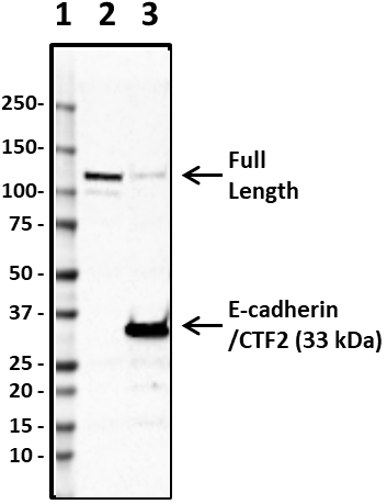
Total cell lysate from HeLa cells (lane 1, 15 µg), Jurkat ce... -
Alexa Fluor® 647 anti-Vimentin

HeLa cells were fixed with 2% paraformaldehyde (PFA) for ten... 
Confocal image of human liver sample acquired using the IBEX... 
Human paraffin-embedded colon tissue slices were prepared wi... 
Confocal image of human jejunum sample acquired using the IB... 
Confocal image of human kidney sample acquired using the IBE... -
Alexa Fluor® 488 anti-Vimentin

HeLa cells were fixed with 4% paraformaldehyde (PFA) for 15 ... 
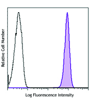
Jurkat cells (filled histogram, positive control) and Daudi ... 
IHC staining of Alexa Fluor® 488 anti-Vimentin (clone O91D3)... -
TotalSeq™-Bn1302 anti-Vimentin


Follow Us